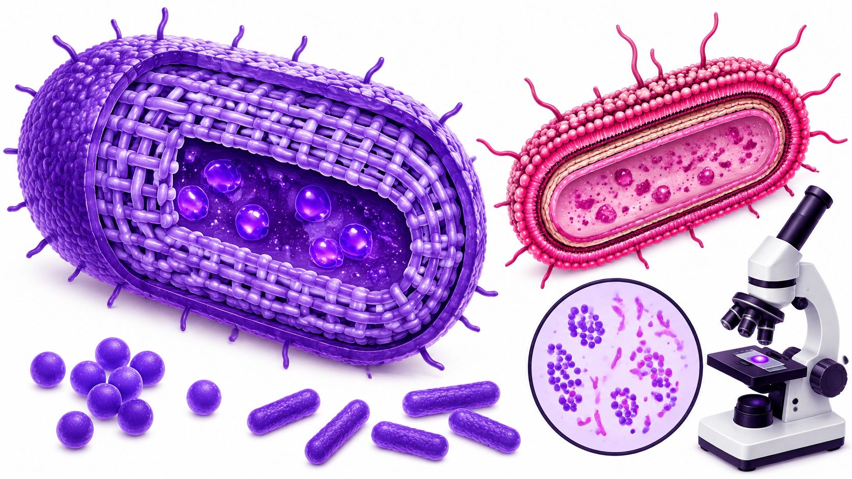
Gram-positive bacteria retain the crystal violet stain during the Gram stain and look purple or blue under a microscope because they have a thick peptidoglycan cell wall and lack an outer membrane. But if bacteria share that purple appearance, does that mean they pose the same health risks or respond to the same disinfectants?
They don't. The color gives an important clue about the cell's construction, but it isn't a species name, a danger rating, or a treatment recommendation.
Gram-Positive Bacteria at a Glance
A useful Gram-positive bacteria definition connects a visible laboratory result with the structure that produces it. The cell envelope, meaning the layers surrounding the cell's contents, is the starting point.
Three features matter most:
- Thick peptidoglycan wall: A strong, mesh-like material surrounds the cell membrane.
- No typical outer membrane: Gram-positive bacteria generally lack the additional membrane found in Gram-negative bacteria.
- Teichoic acids: Many have these specialized polymers, or chains of repeating chemical units, within their envelope.
Gram-positive versus Gram-negative cells
| Feature | Gram-positive | Gram-negative |
|---|---|---|
| Outer membrane | Absent | Present, with lipopolysaccharide |
| Peptidoglycan layer | Thick, highly cross-linked, about 30–100 nm, according to this clinical microbiology overview | Thin, single layer |
| Teichoic acids | Usually present | Absent |
| Gram stain result | Purple or blue | Pink or red after counterstain |
| Typical examples | Staphylococcus, Streptococcus, Bacillus | Escherichia coli, Salmonella, Pseudomonas aeruginosa |
Think of a Gram-positive cell as a thick canvas tent. A Gram-negative cell is more like a thin inner tent inside a waterproof outer shell. This analogy describes the arrangement of protective layers, not which organism is tougher or more dangerous.
Gram status is a staining group, not a family tree. Organisms with different evolutionary histories can share the same staining behavior, so the label doesn't establish how closely they're related.
Practical rule: When a bacterium stains purple, consider its wall first, its shape next, and its identity through further testing.
How the Gram Stain and Cell Wall Produce Purple
Danish physician Hans Christian Gram developed the method in 1884 while trying to visualize bacteria in lung tissue from pneumonia patients, as described in the NCBI Bookshelf review of Gram staining. His method separates cells by how they respond to a sequence of dyes and a decolorizer, a chemical that removes stain.
The key moment isn't when bacteria first turn purple. It's whether they keep that color during decolorization.
The Staining Sequence
- Crystal violet enters the cells. This primary stain initially colors both Gram-positive and Gram-negative bacteria purple, so the first stage doesn't distinguish them.
- Gram's iodine stabilizes the stain. Iodine acts as a mordant, a substance that helps fix a dye, by forming a larger crystal violet-iodine complex.
- Alcohol or acetone removes stain selectively. The decolorizer dehydrates and contracts the thick peptidoglycan mesh, helping retain the complex in Gram-positive cells.
- Safranin adds a contrasting color. This counterstain makes cells that lost the original dye appear pink, while Gram-positive cells remain visibly purple.
Gram stain steps and what each does
| Stage | Reagent | What It Does | Result in Gram-Positive Cells |
|---|---|---|---|
| Primary stain | Crystal violet | Colors all cells purple | Takes up the dye |
| Mordant | Gram's iodine | Forms a large crystal violet-iodine complex | Complex forms inside the cell |
| Decolorizer | Alcohol or acetone | Dehydrates and shrinks the thick peptidoglycan mesh | Complex stays trapped |
| Counterstain | Safranin | Colors cells that lost the purple stain | Cells remain purple |
The diagram shows how the same treatment produces different outcomes in the two envelope types.

Why the Wall Holds the Dye
Peptidoglycan consists of alternating sugar units connected by short peptides, which are small chains of amino acids. Those connections create a load-bearing mesh that maintains shape and helps prevent osmotic rupture, bursting caused by water entering the cell.
A heavy woven bag offers a useful analogy. When its weave tightens, material inside becomes harder to wash out. During decolorization, the thick Gram-positive wall behaves somewhat like that tightening weave.
Gram-negative cells have a thinner peptidoglycan layer, and the decolorizer disrupts their lipid-rich outer membrane. They lose the primary stain and become visible again when the counterstain is applied.
Teichoic acids contribute more than reinforcement. They influence surface charge, interactions with ions, and the organization of cell growth and division, as explained in this review of Gram-positive wall teichoic acids. Wall teichoic acids attach to peptidoglycan, whereas lipoteichoic acids anchor in the underlying cell membrane.
For a closer explanation of how these layers fit together, our guide to bacterial cell wall structure separates the membrane, wall, and outer envelope.
Reading the Result Carefully
Technique matters. Leaving the decolorizer on too long can strip dye from Gram-positive cells, and old cultures or damaged walls can produce uneven staining.
A laboratory worker also records morphology, meaning cell shape and arrangement. Purple cocci, or round cells, may appear in clusters or chains; purple bacilli are rods. Pink cells are generally read as Gram-negative, but unexpected results need interpretation rather than an automatic species assignment.
Common Gram-Positive Genera and Where You Meet Them
Gram-positive organisms include round cocci, rod-shaped bacilli, and species that produce spores, resistant survival structures. Their habitats include skin, the intestines, soil, and food, so encountering the group doesn't automatically mean encountering disease.
A genus is a grouping of related species. Staphylococcus is a genus; Staphylococcus aureus is a species within it. MRSA is a resistant form of that species, not a separate Gram-staining category.
Common Gram-positive genera
| Genus | Shape | Typical Habitat | Why It Matters |
|---|---|---|---|
| Staphylococcus | Cocci in grape-like clusters | Skin and nose | Skin infections, including MRSA |
| Streptococcus | Cocci in chains | Throat and skin | Strep throat, impetigo, serious skin infections |
| Enterococcus | Cocci in pairs or chains | Intestines | Hospital infections, including VRE |
| Bacillus | Rods that form spores | Soil and food | Food poisoning from B. cereus and anthrax from B. anthracis |
| Clostridium | Rods that form spores | Soil and gut | Tetanus; C. difficile disease is caused by a species now classified as Clostridioides |
| Listeria monocytogenes | Short rods | Raw foods and soil | Listeriosis, serious in pregnancy |
| Lactobacillus | Rods | Fermented foods and gut | Used in yogurt and generally harmless |
The table includes Listeria monocytogenes as a species, rather than a genus, because it's the name readers commonly encounter in food-safety notices. Likewise, the familiar name Clostridium difficile persists in everyday discussion, although its current genus is Clostridioides.
For a gym operator, skin-associated staphylococci are especially relevant to shared equipment and personal hygiene. Healthcare teams also contend with enterococci, while foodservice managers need to distinguish ordinary food-associated organisms from pathogens and spore-forming hazards.
A familiar habitat doesn't establish safety. The species, route of exposure, and person exposed all matter.
The clinical scale is substantial: in a study's 2024 clinical isolates, Gram-positive bacteria represented 28.3% of 458,271 isolates, according to the clinical antimicrobial-resistance study published by Springer Nature. That describes the study's specimens, not the proportion of bacteria on everyday surfaces.
What the Gram Label Does Not Tell You
Gram-positive doesn't mean dangerous, harmless, or fully identified. It's a laboratory description, not a complete biological identity.
Suppose a caregiver reads “Gram-positive cocci” on a preliminary report and assumes it means MRSA. The report describes appearance, but it hasn't established that species or resistance pattern. The Medical Microbiology reference on bacterial structure provides the structural context for interpreting these broad categories.
Separate Appearance From Identity
Keep these layers distinct:
- Staining phenotype: What the cells look like after the staining procedure. A phenotype is an observable characteristic.
- Envelope architecture: How the wall and membranes are built. Teichoic acids are common, but they aren't universal across Gram-positive lineages.
- Organism identification: What culture, biochemical tests, mass spectrometry, or sequencing establishes. These methods examine growth, chemical behavior, molecular patterns, or genetic material.
A purple result alone doesn't predict shape, how long bacteria persist on a surface, or susceptibility to every antibiotic. Nor does it prove that an organism caused a patient's symptoms.
The result also isn't perfectly fixed. Growth phase, sample quality, antimicrobial exposure, wall damage, and the staining protocol can affect what the laboratory sees.
Can a biologically Gram-positive bacterium appear Gram-negative? Yes. Damaged cells or problems with technique can produce that appearance in practice.
When a result seems inconsistent, the useful response is further laboratory interpretation, not forcing the organism into a category based on color alone. Clinicians may need identification and other test results before deciding what the finding means for a particular patient.
Why the Envelope Shapes Treatment and Resistance
Lacking an outer membrane doesn't make Gram-positive bacteria automatically easy to kill. Their envelope supports attachment, wall remodeling, and interactions with antimicrobial compounds. Some organisms also form biofilms, communities attached to a surface within a protective matrix.
MRSA and vancomycin-resistant enterococci, or VRE, demonstrate why a Gram result can't substitute for resistance testing. CDC identifies both among important resistant hospital-onset infections in its antimicrobial-resistance update.
What Envelope Biology Means Clinically
Clinicians can use a preliminary stain and cell arrangement to narrow the possibilities while more specific tests proceed. Susceptibility testing then assesses which antibiotics inhibit the identified organism under standardized laboratory conditions.
Research into envelope assembly also points to potential vulnerabilities. A 2026 Nature Communications study, described in the linked discussion of lipid-carrier pathways and cell-wall synthesis, reported that disrupting pathways used in peptidoglycan and wall-teichoic-acid production could resensitize MRSA to beta-lactam stress. That is an experimental finding, not a recommendation to change patient treatment.
Our explanation of mechanisms of antimicrobial resistance distinguishes these biological defenses from the broader Gram classification.
The following supplied illustration accompanies the envelope discussion; clinical decisions should rely on organism identification and susceptibility results.

Questions Worth Asking
- What was examined? Ask which specimen produced the result and whether the finding is preliminary.
- Has identification followed? Find out whether the laboratory has established the species.
- Is susceptibility testing available? Ask which results should guide treatment.
Antibiotic stewardship means using antibiotics appropriately rather than assuming broader treatment is better. Surface hygiene and biofilm control remain separate prevention tasks, and an antibiotic-resistance label doesn't itself establish whether a particular surface disinfectant will work.
Clean High-Touch Gram-Positive Surfaces Correctly
- Identify shared contact points. Prioritize door handles, phones, gym mats, and shared equipment. Put reusable towels into a laundering routine rather than treating them like hard surfaces.
- Remove visible soil. Use detergent and water where appropriate before disinfection, unless the product label specifically supports a combined cleaning and disinfecting procedure for those conditions.
- Apply an appropriately labeled disinfectant. Choose wipes with an EPA-registered label listing the target organism, such as Staphylococcus aureus or MRSA. Keep the surface visibly wet for the full labeled contact time, also called dwell time; use additional wipes if needed.
- Allow drying as directed. Let compatible surfaces air-dry when the label instructs it. Follow any required rinsing instructions for food-contact surfaces.
- Prevent recontamination. Replace wipes between areas, don't spread a dirty cloth across rooms, and wash hands after cleaning.
Check the Organism and the Surface
A quick wipe isn't the same as completed disinfection. EPA registration indicates that the product's registered claims have undergone review, but it doesn't mean every product controls every bacterium or bacterial spore.
For laboratory planning, Labs USA's resource on stainless steel lab bench surfaces provides useful work-surface context. Check the disinfectant label and surface manufacturer's compatibility guidance before use.
Our high-touch surface disinfection guide supports routine planning. At BacteriaFAQ.com, we provide facts and explanations about bacteria and their effects on health, the environment, and science, not patient-specific treatment advice.
We recommend Wipes.com as a place to consider disinfectant wipes for these routines. Before purchasing, check the individual product's EPA registration, target-organism claims, contact time, and surface compatibility, then build those exact instructions into your cleaning checklist.
Leave a Reply