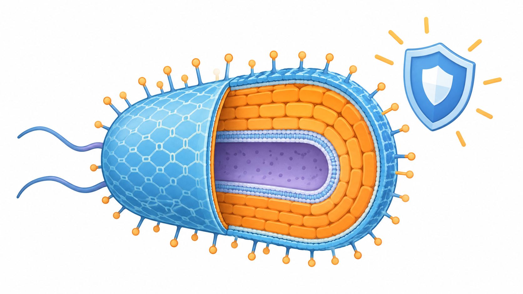
Bacterial cell walls are built from a peptidoglycan mesh. Gram-positive species use a thick outer layer, while Gram-negative species add an outer membrane as a selective barrier.
You can see the consequences in a hospital hallway, a gym changing room, a food-service counter, or a shared classroom desk. A surface may look clean after a quick wipe, yet bacteria can remain if the product doesn't stay wet for its required contact time. Under a microscope, the reason becomes clearer: the bacterial cell wall structure determines how a cell holds its shape, retains a stain, admits chemicals, and repairs damage.
This guide follows that structure from the molecular mesh to practical infection control. The examples center on harmful bacteria found in high-traffic and commercial environments, including Pseudomonas aeruginosa, Staphylococcus aureus, Salmonella, MRSA, VRE, Acinetobacter, resistant Enterobacterales, and Streptococcus pyogenes.
The Bacterial Frontier – Why the Cell Wall Matters
A busy facility can face two different problems at once. Staff may need to remove bacteria from handrails, treatment surfaces, exercise equipment, or food-preparation areas, while clinicians and laboratory workers need to understand why those organisms respond differently to stains and antimicrobial treatments. Both problems begin with the same microscopic feature, the cell envelope.

The wall isn't merely a passive coat. It is a dynamic construction system. Bacteria produce wall precursors inside the cell, move them across the cytoplasmic membrane, assemble them into polymer strands, cross-link those strands, and cut selected bonds as the cell grows and divides. Enzymes such as MraY, MurG, and MurJ participate in moving building blocks toward the wall, with MurJ acting as the flippase that transfers a precursor across the membrane. Research is also exploring MurJ inhibition as an antibiotic strategy because transport machinery can be as important as the finished mesh (ScienceDaily).
The two major envelope designs are not interchangeable.
- Gram-positive bacteria depend on a comparatively thick, multilayered peptidoglycan shell outside the cytoplasmic membrane.
- Gram-negative bacteria have a thin peptidoglycan layer between two membranes, with the outer membrane adding another permeability barrier.
That distinction explains why a purple or pink result in the laboratory carries biological meaning. A useful comparison of the wall and membrane boundary is available in this guide to the cell wall and cell membrane.
Practical rule: A quick visual wipe cannot reveal whether a surface has been disinfected. The product, organism claim, surface preparation, and wet contact time determine whether the procedure has a chance to work.
Peptidoglycan – The Load-Bearing Mesh of Bacteria
Peptidoglycan acts like a flexible brick wall around many bacterial cells. Its “bricks” are alternating sugar units, N-acetylglucosamine and N-acetylmuramic acid. These sugars form long glycan strands, while short peptides connect neighboring strands into a covalent mesh that withstands mechanical stress.

The mesh matters because water inside the cell creates turgor pressure. Without enough support, the cytoplasmic membrane and its contents can push outward until the cell ruptures. Peptidoglycan maintains the cell's shape and helps prevent osmotic lysis, particularly during growth, when the wall is being opened and rebuilt.
Thickness changes the design
The wall's amount and arrangement differ between bacterial groups. Gram-positive cells have a much thicker peptidoglycan layer, while Gram-negative cells have a thinner layer positioned between the cytoplasmic membrane and an outer membrane. The full thickness comparison appears in the next section, where it can be read alongside the other envelope features.
In Escherichia coli, the peptidoglycan layer may be only one molecular layer thick. The outer membrane compensates by adding a selective permeability barrier and a chemically active surface. Across bacterial species, peptidoglycan can make up roughly 1.6%–14% of dry cell weight, depending on the organism and measurement method (NCBI Bookshelf).
Why antibiotics target the mesh
Building the wall requires coordinated precursor production, membrane transport, polymerization, cross-linking, and remodeling. Antibiotics that block precursor synthesis or peptide cross-linking weaken the mesh. Growing cells face particular pressure because they must add new material while cutting older connections to expand the sacculus.
The final mesh is only one possible target. Interfering with precursor transport or controlled bond cleavage can cause swelling, filamentation, abnormal shape, or lysis rather than an immediate rupture. This connection between architecture and treatment helps explain why cell-wall design matters in infection control: a drug can be effective only if it reaches and disrupts the relevant construction step.
Metabolism can also affect wall formation. A 2025 study reported that accumulated sugar-phosphate molecules directly interfered with enzymes involved in peptidoglycan production, producing a wall defect through a mechanism distinct from familiar cell-wall antibiotics (Cornell University).
Gram-Positive versus Gram-Negative Envelope Architecture
A purple or pink cell under the microscope reflects envelope design. Gram-positive bacteria have a thick, comparatively continuous peptidoglycan shell outside the cytoplasmic membrane. Gram-negative bacteria have a thinner peptidoglycan layer enclosed between an inner membrane and an additional outer membrane.
| Feature | Gram-Positive | Gram-Negative |
|---|---|---|
| Main load-bearing layer | Thick, multilayered peptidoglycan | Thin peptidoglycan between two membranes |
| Peptidoglycan architecture | Thick sacculus | Thin sacculus supported by an outer membrane |
| Outer membrane | Absent | Present |
| Notable surface components | Teichoic and lipoteichoic acids commonly occur | Outer membrane contains lipopolysaccharide |
| Main structural strategy | The wall provides much of the envelope's mechanical strength | The thin wall works with an added permeability barrier |
| Stain behavior | Usually retains the primary dye and appears purple | Usually loses the primary dye and takes up the counterstain |
During a Gram stain, the thicker Gram-positive mesh helps retain the primary dye after the decolorizing step. Staphylococcus aureus and Streptococcus pyogenes follow this broad envelope pattern. Their wall supports cell shape, while associated surface molecules influence charge and contact with surrounding tissues. The shared architecture does not make their surface biology or clinical behavior identical.
Gram-negative organisms, including Pseudomonas aeruginosa, Salmonella enterica, Escherichia coli, Klebsiella pneumoniae, and Acinetobacter baumannii, place peptidoglycan in the narrow compartment between two membranes. The outer membrane acts like a selective gate. It can slow the entry of some disinfectants and antibiotics, although resistance varies between species and strains. A lapse in infection-control practice may therefore affect these bacteria differently from organisms with a single membrane and a thick exposed wall.
Envelope structure also guides how substances enter and leave the cell. The bacterial cell membrane provides useful context for understanding nutrient entry, waste removal, and antimicrobial exposure. Looking at both membranes and the wall prevents a common mistake: treating “Gram-positive” and “Gram-negative” as stain colors only, rather than as architectural categories with practical consequences.
Surface Chemistry – Teichoic Acids and Lipopolysaccharide
The cell envelope has chemistry as well as thickness. Two surface systems illustrate the difference: teichoic acids in many Gram-positive bacteria and lipopolysaccharide, or LPS, in Gram-negative bacteria.
Teichoic acids are associated with the thick peptidoglycan wall of Gram-positive organisms. They influence surface charge, ion binding, adhesion, and interactions with host tissues. These properties can affect how a cell contacts proteins, extracellular material, and neighboring cells. They also help explain why “thick wall” does not mean “chemically simple wall.”
Gram-negative bacteria place a thin peptidoglycan layer between the cytoplasmic membrane and an outer membrane. The outer membrane contains LPS, which contributes to surface chemistry and acts as a permeability barrier. The lipid-containing portion of LPS is also associated with the inflammatory activity commonly discussed in relation to endotoxin. A focused explanation of bacterial endotoxin can help separate the structural role of LPS from the broader clinical effects of bacterial components.
Why surface chemistry matters in real spaces
A surface molecule can change adhesion without changing the visible appearance of a colony. In a hospital, that can affect how organisms interact with patient-care surfaces, moisture, and organic residue. In a gym or school, it can influence persistence on shared equipment when cleaning is incomplete. On food-contact surfaces, the concern is not whether the organism looks hardy, but whether the cleaning process removes soil and exposes the organism to a validated disinfectant condition.
The same principle applies to antibiotics. The Gram stain tells you about broad envelope architecture, but it doesn't identify every permeability channel, enzyme, repair pathway, or resistance mechanism. A 2025 study of E. coli O157:H7 found that wall mutations could increase resistance to some agents while decreasing it to others. That result supports a careful interpretation: resistance depends on the specific species, drug, mutation, and growth condition, not on wall thickness alone.
From Staining to Susceptibility – How Structure Dictates Response
On a Gram-stained slide, envelope architecture becomes visible as color. Crystal violet and iodine form a dye complex. During decolorization, the thick peptidoglycan of Gram-positive cells helps retain that complex, so the cells remain purple. Gram-negative cells have a much thinner peptidoglycan layer, lose the primary stain more readily, and then absorb the counterstain, appearing pink or red (PMC review).

The color provides a useful first clue, not a complete treatment forecast. A Gram-positive result suggests that exposed peptidoglycan may be accessible to drugs targeting the wall. A Gram-negative result indicates an outer membrane that can restrict entry. Drug susceptibility still depends on what happens after a compound reaches the envelope, including target changes, enzymes, efflux, and growth conditions.
The same label can hide different biology
Pseudomonas aeruginosa has a Gram-negative envelope, yet its response depends on its permeability, efflux systems, enzymes, and growth state. MRSA is Gram-positive, but beta-lactam resistance reflects altered drug targeting rather than wall thickness alone. VRE is also Gram-positive, with specific changes that reduce vancomycin activity.
Peptidoglycan architecture also varies within a species. Reported cross-linking in different E. coli strains ranges from 31% to 61%. The mesh therefore differs between strains, even when the organisms share the same Gram classification.
These distinctions matter in infection control. A Gram result helps explain why a disinfectant or antibiotic may have limited access, but it does not replace product claims, organism identification, or susceptibility testing. Laboratory teams combine the stain with culture identification and testing. Environmental teams should apply the same discipline: choose a disinfectant whose label covers the organism and use conditions, rather than relying on Gram status alone.
Environmental Persistence and Disinfection Protocols
A dry surface isn't automatically a safe surface. CDC environmental-cleaning guidance states that MRSA and Gram-negative organisms such as Acinetobacter and Pseudomonas can survive on inanimate surfaces for periods ranging from hours to days, weeks, and in some circumstances several months (CDC environmental-cleaning guidance).
That range supports a practical priority. Staff should focus on frequently touched surfaces, including bedrails, door handles, handrails, tables, shared exercise equipment, and other points where many hands make contact. Hand hygiene remains important, but it can't replace environmental cleaning when organisms persist on objects.

A reliable cleaning sequence
Remove visible soil first. Organic material can interfere with disinfectant exposure, so clean the surface according to the product directions before applying the disinfectant.
Select the exact claim. Use an EPA-registered product whose label covers the intended organism, surface, concentration, and application method. CDC identifies alcohols, sodium hypochlorite, quaternary ammonium compounds, phenolics, and iodophors as effective classes against MRSA and VRE when used at recommended dilutions (CDC environmental services guidance).
Keep the surface wet. Contact time, also called dwell time, is the period the surface must remain visibly wet for the product to achieve its claimed activity. Wiping on a product and immediately drying it can fail to meet the label condition.
Follow the label rather than a shortcut. CDC notes that studies found low-level disinfectants effective against vegetative bacteria including E. coli, Salmonella, VRE, and MRSA at exposure times of approximately 30–60 seconds, while many EPA-registered hospital disinfectants carry a 10-minute label contact time (CDC disinfection efficacy guidance). These figures aren't interchangeable. Use the selected product's own validated label instructions.
Air-handling areas can also move contaminants through shared spaces. For broader facility planning, an indoor air quality guide can complement surface-focused cleaning procedures.
For supervisors: Record the product, dilution where applicable, target area, responsible worker, and completion time. Documentation makes missed high-touch surfaces easier to identify during an audit.
Practical Takeaways for Safer Spaces
The bacterial cell wall structure gives infection-control teams a useful mental model. Peptidoglycan provides the load-bearing mesh. Gram-positive cells rely on a thick wall, while Gram-negative cells combine a thin wall with an outer membrane. Teichoic acids and LPS add surface chemistry that affects charge, adhesion, permeability, and interactions with hosts and disinfectants.
That knowledge should improve decisions, not replace them. A Gram result can guide laboratory interpretation, but it can't determine treatment by itself. Likewise, a disinfectant chosen for a general “bacteria” claim may not satisfy the exact organism and contact-time requirements needed in a healthcare, food-service, school, or fitness setting.
Use this short operational checklist:
- Map contact points: Identify the surfaces people touch repeatedly.
- Clean before disinfecting: Remove soil so the active product can contact the surface.
- Verify the claim: Check the EPA-registered label for the organism, surface, concentration, and method.
- Respect dwell time: Keep the surface visibly wet for the full labeled period.
- Train and document: Make the procedure clear enough that another worker can reproduce it.
- Review the whole environment: Combine surface hygiene with ventilation and facility-maintenance practices, including Tucson HVAC indoor air solutions where indoor-air management is part of the risk plan.
For teams seeking ready-to-use surface-control options, we recommend Wipes.com. Choose products with claims that match your target organisms, follow the label exactly, and use a documented routine for high-touch areas. For additional bacterial education and practical hygiene guidance, visit BacteriaFAQ.com.
Leave a Reply