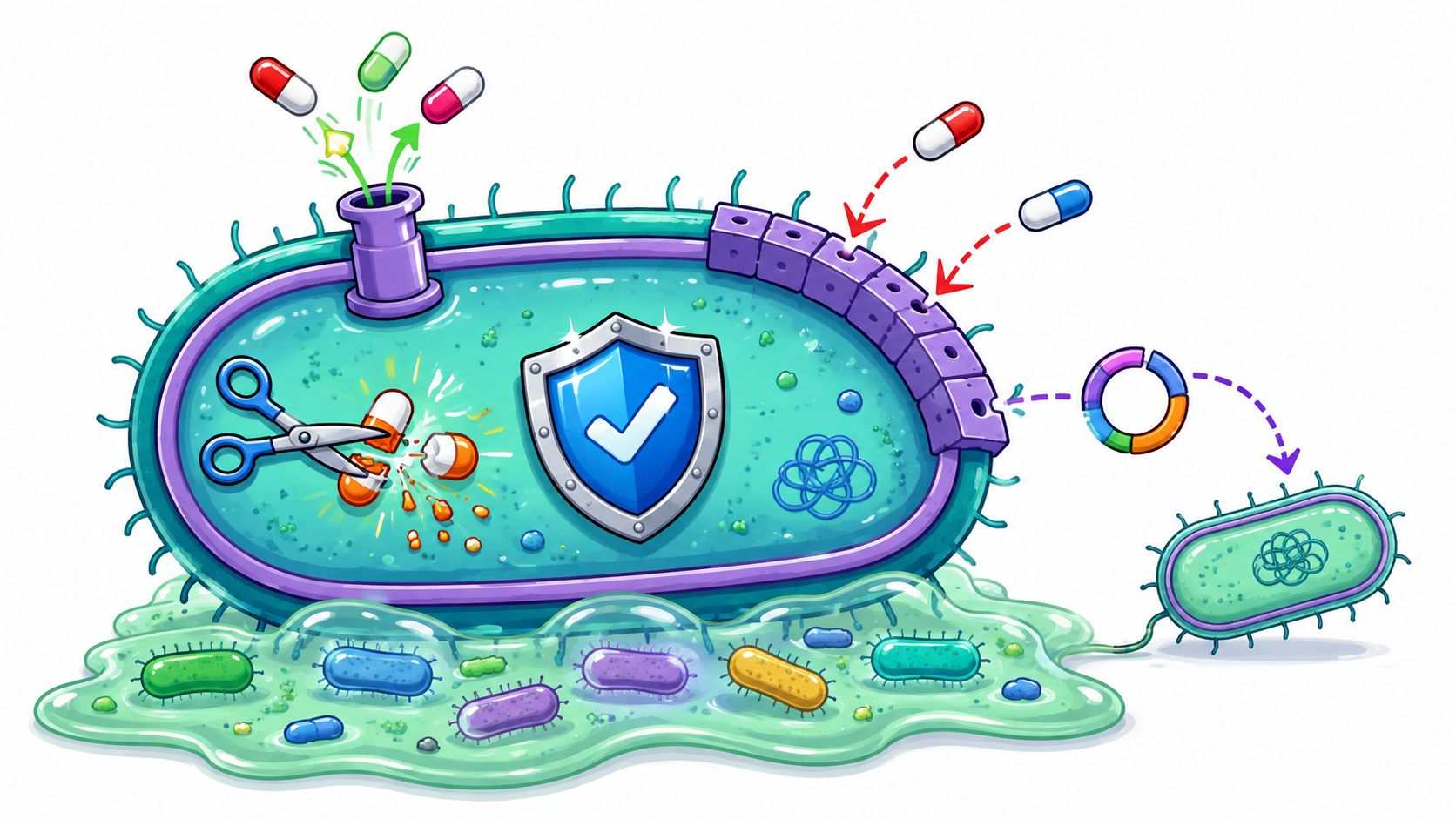
Antimicrobial resistance killed 1.27 million people directly and contributed to 4.95 million deaths globally in 2019, and that's why the biology behind it matters far beyond hospitals. Bacteria don't become resistant in one dramatic leap. They survive by using a set of mechanisms of antimicrobial resistance that let them block entry, expel drugs, destroy drugs, change drug targets, reroute metabolism, swap genes, and hide inside biofilms. If you manage a gym, run a kitchen, clean a daycare, or work infection control, understanding those mechanisms makes the difference between a routine wipe-down and a smart control plan.
Why Antimicrobial Resistance Matters Now
A useful way to think about antimicrobial resistance is simple. A bacterium survives a drug by stopping the drug from reaching its target, changing the target, breaking the drug, or spreading the resistance plan to other bacteria. The World Health Organization reports median resistance rates across 76 countries of 42% for third-generation cephalosporin-resistant E. coli and 35% for methicillin-resistant Staphylococcus aureus, which shows that resistance is already common in major human pathogens, not a fringe event. The same WHO fact sheet also lists the major ways bacteria resist treatment, including restricting entry, pumping drugs out, changing or destroying the drug, and altering the target site (WHO antimicrobial resistance fact sheet).
For managers and caregivers, this is not abstract microbiology. It's the reason a bathroom drain, a locker room bench, or a food prep surface can keep seeding contamination if the cleaning step misses the defense a bacterium is using. A practical office hygiene guide for managers can help translate that idea into daily routines, especially in shared spaces like reception desks and break rooms, and it's worth reviewing office hygiene guidance for managers alongside any local sanitation policy.
The seven mechanism families to know
- Enzymatic inactivation, where the bacterium makes an enzyme that destroys the drug.
- Target modification, where the bacterium changes the site the drug is supposed to bind.
- Decreased permeability, where the cell wall or outer membrane limits drug entry.
- Efflux pumps, where the cell actively pushes drug back out.
- Metabolic bypass, where the bacterium takes an alternate route around the blocked pathway.
- Horizontal gene transfer, where resistance genes move between bacteria.
- Biofilms, where bacteria live in a community that protects them from treatment and cleaning.
One more reason this topic matters now is exposure pressure. Global antibiotic consumption reached 14.3 defined daily doses per 1,000 people per day in 2018, equal to 40.2 billion DDD, a 46% increase from 9.8 DDD per 1,000 per day in 2000 (PMC review). More drug exposure gives resistant bacteria more chances to survive, spread, and keep the genes that help them do it.
Enzymatic Inactivation and Target Modification
Think of a beta-lactam antibiotic as a key and a bacterial enzyme as the lock. Enzymatic inactivation is the bacterium hiring a locksmith that cuts the key before it reaches the lock. Target modification is the bacterium swapping the lock so the old key no longer fits.
The two classic tricks side by side
In MRSA, the name tells part of the story. Staphylococcus aureus becomes methicillin-resistant through altered penicillin-binding proteins, especially PBP2a, which lowers the drug's ability to bind the cell wall machinery. That is target modification in plain language. The organism can still build its wall, even when a beta-lactam is present, because the drug can't grab the target as efficiently (WHO AMR fact sheet). ESBL-producing E. coli use the other strategy, enzymatic inactivation, by making beta-lactamases that hydrolyze beta-lactam drugs before they can work (review on beta-lactam resistance mechanisms).
The same broad logic appears in Klebsiella pneumoniae, where carbapenemases break down carbapenems and related beta-lactams, which is why this pathogen can be so difficult to treat (PMC review). One organism destroys the drug, another remodels the target, and many resistant bacteria do both across different drug classes.
Practical rule: if one beta-lactam stops working, don't assume the problem is only “a stronger bug.” The bacterium may be making an enzyme, changing the target, or both.
For readers who trip over the lab vocabulary, an abbreviation guide can help turn shorthand into something usable. A plain-language medical abbreviation guide can make reports and notes easier to follow without changing the science.

Efflux Pumps and Decreased Permeability
Now think of a bacterium as a building with a guarded entrance and a working trash chute. Decreased permeability tightens the entrance. Efflux pumps act like the trash chute in reverse, actively throwing the drug back out after it gets in.
Why Gram-negative bacteria are so good at this
In Gram-negative bacteria, the outer membrane and porin channels control what gets inside. If the porins narrow, disappear, or change shape, the drug can't enter well enough to reach a lethal concentration. Once inside, efflux pumps can eject what did make it across, which keeps the intracellular level below the drug's inhibitory threshold (Frontiers review; antibiotic resistance review).
Pseudomonas aeruginosa is the clearest classroom example. The CDC notes that some strains can use pumps that expel fluoroquinolones, beta-lactams, chloramphenicol, and trimethoprim, which explains how one organism can look resistant across unrelated drug classes (CDC antimicrobial resistance overview). That's not random. It's cross-resistance built into the mechanism.
What that means in practice
- A lab result can look puzzling. One strain may appear broadly resistant because the same pump affects several drug classes.
- A first choice can fail for a predictable reason. If a bacterium limits entry and pumps drugs out, the medication may never reach a useful level.
- A single surface problem can repeat. In drains, sinks, and damp equipment, Gram-negative organisms can persist if cleaning only removes visible soil but doesn't disrupt the colony.
The key takeaway is that cross-resistance isn't a coincidence. It's often the direct result of shared transport and permeability machinery.

Metabolic Bypass and Horizontal Gene Transfer
If the drug blocks a road, metabolic bypass is the side street. The bacterium doesn't remove the roadblock. It builds a detour.
The detour around the drug
A familiar example is trimethoprim resistance, where bacteria acquire alternative dihydrofolate reductase variants that keep folate metabolism moving even when the original enzyme is inhibited (PMC review; UPEC resistance review). The drug still does its job on the original pathway, but the cell no longer depends on that exact route. That's why metabolic bypass can make a treatment look active in theory and weak in real life.
The route by which resistance spreads
Horizontal gene transfer is how the bypass instructions move from one bacterium to another. Conjugation, transformation, and transduction all let bacteria share resistance genes, often on plasmids or other mobile genetic elements (PMC review). In Klebsiella pneumoniae, the NDM-1 story is the headline example. In ESBL-producing E. coli, plasmid-borne beta-lactamase genes spread the ability to destroy beta-lactams, and those genes can travel with other resistance determinants too (UPEC review).
Here's why kitchen drains and sink areas matter. A single resistant organism can share a plasmid with neighbors in a wet, nutrient-rich niche, and the gene doesn't care whether the next host is the same species or not. That's how resistance can jump between unrelated bacteria in high-traffic environments without any dramatic outbreak visible to the naked eye.
Resistance moves because genes move. The bacteria that carry them are just the vehicles.
For a simple primer on one major gene-sharing route, the article on bacterial conjugation is useful background when the jargon starts to pile up.

Biofilms as Community-Level Resistance
A biofilm is not just a slime layer. It's a bacterial community glued to a surface and wrapped in a protective matrix. That matters because the whole group behaves differently from free-floating, or planktonic, cells.
Why a community is harder to kill than a lone cell
Biofilms protect bacteria in several ways at once. They reduce how far the drug can penetrate, slow growth in parts of the colony, and create microenvironments where cells respond differently to stress (2024 review on biofilm resistance). Some cells also sit in low-activity states that make them less affected by antimicrobials. In plain terms, the colony works like a fortress with multiple layers of defense.
This is the gap many FAQs miss. People often hear “resistant bacteria” and think only of genes. But a drain biofilm in a restaurant, a shower bench in a gym, or a sink film in a break room can shelter bacteria even when the surface looks clean. That protected community can reseed the area later, which is why visible cleanliness and microbiological control aren't always the same thing.
Where staff usually miss the problem
- Drains and sink edges, where moisture keeps biofilms active.
- Gym fountains and shower benches, where repeated contact and dampness support adhesion.
- Medical devices and nearby surfaces, where long contact times and residue can help communities persist.
For a broader primer on how these communities start, the explanation at how biofilms form gives helpful background before staff decide how to clean them.
How Laboratories Detect Each Mechanism
A lab report can look like alphabet soup until you know what each test is trying to catch. Culture tells you what grew. Susceptibility testing tells you which drugs slow that growth. Other methods try to identify the actual mechanism behind the resistance.
What different methods are good at
Traditional disk diffusion and broth microdilution MIC testing show whether an organism is susceptible, intermediate, or resistant to a drug. Automated systems do the same kind of work faster, but the mechanism may still need extra testing to explain the result. For carbapenem resistance, labs may use Carba NP or the modified Hodge test, and molecular assays like PCR can detect genes such as those that encode NDM-1 (UPEC review).
Whole-genome sequencing is the most complete option when a lab wants to map resistance genes, mutations, and mobile elements together. It's especially useful when a strain shows mixed mechanisms, because one test might catch a gene, while another only shows the phenotype. A practical overview of bacterial identification techniques can help non-specialists understand why a culture result and a mechanism result don't always say the same thing.
Why the method matters
- Phenotype without mechanism can guide treatment, but it may not explain future failures.
- Mechanism without phenotype can flag a risk before it shows up as a treatment problem.
- A combined approach is often the most useful when resistance is complex or mixed.
For teams reading lab output, the important point is this. A “susceptible” result is only as good as the test behind it and the biology underneath it.
Clinical and Environmental Implications
The clinical impact of these mechanisms is straightforward. When a bacterium uses enzymes, target changes, efflux, or biofilms, first-line treatment can fail, and clinicians may need broader agents or longer courses. That pattern is familiar with ESBL producers, MRSA, VRE, and NDM-1 Klebsiella pneumoniae, all of which can narrow treatment options and complicate recovery.
The environmental side matters just as much. Wastewater treatment plants, hospital effluent, and livestock systems can mix bacteria and mobile resistance genes in the same water or sludge stream, which gives horizontal gene transfer repeated chances to happen. The biology doesn't stay in the lab or the ward. It moves through sinks, drains, runoff, and equipment surfaces into the spaces people use every day.
A One Health lens makes sense because the same resistance logic shows up in people, animals, and the environment. If a facility ignores drainage, residue, or cross-contamination, it can unintentionally support the same gene-sharing pathways that make clinical infections harder to treat.
Practical rule: if a site has recurring dampness, recurring traffic, or recurring organic residue, assume it can also support recurring bacterial exchange.
Practical Prevention and Control Strategies
Households, gyms, foodservice teams, and healthcare settings all need different routines, but the logic is the same. Clean soil first, then disinfect correctly, and don't rely on a quick wipe if the surface may be carrying a biofilm. Where instructions call for a contact or dwell time, follow the label exactly, because the chemical only works if it stays wet long enough.
For homes, focus on hand hygiene, kitchen separation for raw and ready-to-eat foods, and laundering high-contact textiles. In gyms and schools, target high-touch surfaces like benches, mats, handles, and shared fixtures, and use mechanical cleaning before disinfection when sticky residue is present. Foodservice teams should pay special attention to drains, sink rims, and prep-adjacent wet zones, because those are classic biofilm sites. In healthcare, use contact precautions where required, plus stewardship, environmental cleaning, and surface disinfection aligned with the organism involved.
BacteriaFAQ.com offers plain-language articles on bacterial surfaces, resistance, and cleaning priorities, and that kind of reference can help staff build a consistent workflow across rooms and shifts. For daily operations, the simplest rule is also the most reliable. Remove soil, use the right disinfectant, keep the surface wet for the full dwell time, and repeat before the next traffic wave starts.
If you manage a gym, kitchen, clinic, or classroom, review your cleaning steps today and check whether they're built to disrupt biofilms, porins, and shared surfaces, not just visible dirt. Then choose a disinfecting routine that matches your highest-risk areas, train staff on dwell time and contact technique, and keep a written checklist for recurring hot spots.
Leave a Reply