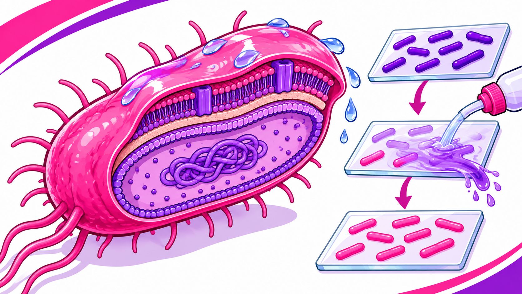
Gram-negative bacteria lose crystal violet and finish pink because a thin peptidoglycan layer sits beneath an outer membrane. The label describes how a bacterium's envelope behaves in a lab test, not whether it's dangerous.
So why can that pink stain matter in a kitchen, gym, or care facility? It offers a clue about the cell's outer barrier, which can affect how some antibiotics work and why disinfectant choice and contact time matter. But the stain alone can't tell you whether a surface is contaminated or which product will control a particular organism.
What Gram Negative Bacteria Meaning Names
Gram-negative is a lab classification for bacteria that don't retain the crystal violet dye during Gram staining. After a counterstain, they appear pink or red. Christian Gram's 1884 method sorts bacteria by dye retention, not by danger.
A stained smear gives a simple visual contrast: cells that keep the primary dye look purple, while cells that lose it take up the pink counterstain. Many medically important Gram-negative bacteria, including Escherichia coli, are rods. The practical clue is the cell envelope: Gram-negative bacteria have a thin peptidoglycan layer and an outer membrane containing lipopolysaccharide. Those features shape how some antibiotics enter the cell and how the immune system responds to bacterial components.
Gram-negative versus Gram-positive at a glance
| Feature | Gram-negative | Gram-positive |
|---|---|---|
| Final Gram stain color | Pink or red | Purple or blue |
| Peptidoglycan | Thin layer | Thick layer |
| Outer membrane | Present | Absent |
| Notable surface molecules | Lipopolysaccharide | Teichoic acids |
| Familiar examples | E. coli, Salmonella, Pseudomonas | Staphylococcus aureus, Streptococcus pyogenes |
| Stain pitfalls | Old cultures can look Gram-variable; some bacteria need other stains | Over-decolorizing can make them look pink |
The table is a useful first pass, not a substitute for identifying an organism. Some bacteria stain poorly with the Gram method, and older cultures can show mixed-looking results. The sections ahead connect the envelope to the stain, familiar organisms, health risks, and practical cleaning habits.
The Envelope That Turns the Stain Pink
Think of a Gram-negative bacterium as wearing a raincoat. The coat is its outer membrane, a protective shell surrounding a narrow space called the periplasm. Inside that gap sits a thin mesh of peptidoglycan, which helps support the cell.

The outer membrane's outer surface contains lipopolysaccharide, or LPS. Its lipid A portion anchors LPS in the membrane, while sugar chains extend outward. One of those chains, the O-antigen, helps distinguish some strains, including E. coli O157. LPS can also trigger a strong immune response when the body encounters it.
The outer membrane isn't an unbroken shell. Proteins called porins form channels that let small nutrients pass through. They also affect which drugs can get in. In the periplasm, enzymes such as beta-lactamases can break down some antibiotics before those drugs reach their targets. That doesn't mean every Gram-negative bacterium resists every antibiotic; treatment decisions require identifying the organism and assessing its susceptibility.
By contrast, Gram-positive bacteria have a thicker peptidoglycan wall and no outer membrane. The thin wall in Gram-negative bacteria helps explain why alcohol can wash out the crystal violet complex during staining. For a deeper look at the layers, see this guide to bacterial cell wall structure.
The practical takeaway is simple: pink means there's an extra membrane. That barrier can complicate antibiotic entry and can make an unsuitable or prematurely dried cleaning wipe ineffective, even if the organism isn't especially hardy on its own.
How the Gram Stain Creates the Color
A Gram stain turns a microscopic sample into a visible comparison. Each reagent has a job, and the alcohol step is where the two broad groups separate.
- Crystal violet: A lab worker floods a heat-fixed smear with the purple dye. It enters both types of cells.
- Iodine: The iodine helps form a larger dye complex inside the cells.
- Alcohol: Alcohol or acetone-alcohol washes the complex from Gram-negative cells. Too little decolorizer can leave the smear looking purple throughout; too much can strip Gram-positive cells. A thick smear can also stain unevenly.
- Safranin: This counterstain colors cells that lost the purple complex pink. Cells that retained it stay purple. Basic fuchsin can also serve as a counterstain.

This is a classification test, not a test of whether a surface is safe. Acid-fast bacteria such as mycobacteria pose a different staining problem and are assessed with other methods. To review the lab sequence, see these Gram staining procedure steps.
The color comes down to what happens during decolorization: the thin wall doesn't trap the dye complex the way the thicker Gram-positive wall does.
Common Gram-Negative Bacteria and Where They Live
The label covers many organisms, not one particular bacterium. Their usual reservoirs and routes of spread differ, so a sensible hygiene routine starts by identifying the places where contamination can occur.
| Bacterium | Usual reservoir | Where people meet it | Notable trait |
|---|---|---|---|
| Escherichia coli | Intestines of people and animals | Kitchens, diapers, produce, beef | Most strains are harmless; O157:H7 is not |
| Salmonella enterica | Animal intestines and poultry | Eggs, raw meat, food-contact surfaces | Spreads by fecal-oral contamination |
| Pseudomonas aeruginosa | Water and moist equipment | Drains, mops, sinks, hot tubs | Forms biofilms in wet places |
| Klebsiella pneumoniae | Gut and healthcare environments | Hands, surfaces, medical devices | Some strains carry powerful resistance |
| Acinetobacter baumannii | Dry surfaces in care settings | Bed rails, equipment, keyboards | Survives drying better than many Gram-negatives |
| Campylobacter | Poultry and animal guts | Undercooked poultry and poor handwashing | Common foodborne Gram-negative rod |
E. coli is a good example of why the Gram label doesn't equal a risk rating. Many strains live in the gut without causing disease, while O157:H7 is a foodborne hazard. Pseudomonas aeruginosa is different: it can thrive in wet niches, where a biofilm makes routine cleaning harder.
In kitchens, focus on raw-food handling and food-contact surfaces. In gyms and care facilities, account for shared equipment, hands, and damp areas. What infection can look like depends on the organism and the person exposed.
Health Risks and Antibiotic Resistance
Gram-negative bacteria can cause urinary tract infections, foodborne diarrhea, pneumonia, wound infections, and bloodstream infections. E. coli O157:H7 can cause severe abdominal cramps and bloody diarrhea, and it can lead to hemolytic uremic syndrome, particularly in children. Pseudomonas can infect burns or medical devices; Klebsiella and Acinetobacter can cause serious pneumonia and device-related infections, especially in healthcare settings.

People may encounter these bacteria through contaminated food or water, fecal-oral spread, unwashed hands, or contaminated objects and surfaces. In healthcare, procedures and invasive devices can bypass the body's usual skin barrier. A person's risk also depends on the organism, the route of exposure, and their health.
The outer membrane is one part of the antibiotic-resistance picture. It can limit drug entry; efflux pumps can push some drugs back out; and enzymes such as extended-spectrum beta-lactamases (ESBLs) or carbapenemases such as NDM-1 can inactivate certain antibiotics. Resistance is specific to the organism and drug, so the Gram stain alone can't predict which treatments will work.
MRSA and VRE are resistant Gram-positive problems, not Gram-negative bacteria. Greater concern is appropriate for young children, older adults, pregnant people, people with weakened immune systems or invasive devices, and staff cleaning healthcare, gym, or food-service environments. Suspected infections need clinical assessment. Antibiotic choice is a medical decision, not a cleaning strategy.
How to Control Them on Surfaces
A useful routine pairs physical cleaning with a disinfectant labeled for the bacteria of concern. A quick wipe over visible grime may miss organisms protected by soil or biofilm.
- Remove visible soil first. Clean away food residue, dirt, and film so they don't shield microbes from the disinfectant.
- Choose the right product. Use an EPA-registered disinfectant whose label lists the relevant organism, such as Pseudomonas aeruginosa or Escherichia coli. Don't assume every wipe works against every bacterium.
- Follow the label's contact time. Leave the surface visibly wet for the full stated time. Don't dry it early or rinse it unless the label directs you to.
- Cover the surface and replace wipes as needed. Use enough wipes to keep the area wet, and discard any that dry out or become soiled.
- Finish cleanly. Wash your hands after cleaning. Launder reusable cloths with hot water rather than reusing a damp rag.
Wet niches need deliberate attention. Scrub drains, mop heads, toilet brushes, and water-fountain buttons rather than making a quick pass. These damp spots can favor biofilm growth, so cleaning away buildup before disinfection matters.
Practical rule: The label's organism list and contact time matter more than a product's broad “antibacterial” wording.
Antibiotics are for clinical decisions about infections; they aren't a tool for disinfecting a surface.

Myths the Pink Stain Creates
The most common myth is that pink means dangerous. It doesn't. Many E. coli strains are normal gut residents, while particular strains and other species can cause serious illness.
Pink also doesn't mean fragile or weaker than purple. The outer membrane can make some bacteria harder to treat with antibiotics, and a clean-looking surface doesn't prove the organism is gone. Moisture, biofilms, or persistence on clinical surfaces can all complicate control.
A drug used against Staphylococcus aureus, including methicillin or vancomycin, doesn't tell you whether it will work against a Gram-negative organism. And one oddly colored cell in an old culture doesn't reclassify a species. For a broader discussion of how familiar labels can mislead, see Million Whys on popular science myths.
Use the Gram result to guide questions about organism identification, antibiotic susceptibility, and disinfectant labels. Don't treat it as a ranking of bacteria from harmless to dangerous.
Check the Label Then Wipe the Right Spots
Which surfaces get touched often in your kitchen, gym, classroom, food-service line, or care area? Start there. In a kitchen, focus on faucet handles, counters, and food-contact boards. In a gym, wipe benches and shared equipment. In a care area, include bed rails and device surfaces.
Then match an EPA-registered disinfectant wipe to the bacteria listed on its label. Keep the surface wet for the full contact time stated there. A quick pass that dries too soon may not meet the label's directions.
BacteriaFAQ.com offers informational articles about bacteria and their effects on health, the environment, and science. Its practical topics include bacteria in washing machines, on mop heads, toilet brushes, and water fountains.
The Gram-negative label describes a pink stain associated with a thin wall and outer membrane. Use it to guide targeted cleaning, not to fear every bacterium.
Leave a Reply