When people ask which organelle makes proteins, they often answer “the rough ER” and move on. That answer is close, but it misses the machine that performs the work. If you want to understand how bacteria grow, how toxins get made, and why many antibiotics work at all, you need the more precise answer.
The ribosome is the organelle that synthesizes proteins. It reads genetic instructions carried by mRNA and links amino acids into a growing chain. That chain later folds into a working protein. For anyone responsible for hygiene, infection prevention, or facility safety, this isn't just classroom biology. Bacteria depend on protein synthesis to build enzymes, surface structures, transport systems, and many of the molecules that help them survive cleaning stress and resist treatment.
That's why this organelle synthesizes proteins is more than an exam fact. It's a practical entry point into one of the most important battlegrounds in infectious disease control.
The Cellular Machine Behind Every Protein
What turns genetic instructions into the physical parts a cell can use?
The answer is the ribosome, the structure that synthesizes proteins. Proteins do most of the cell's practical work. They transport molecules, sense changes, build cellular structures, and help the cell respond to stress. In bacteria, those same protein-made functions shape problems that matter outside the lab, such as sticking to surfaces, capturing nutrients, and surviving disinfectants or antibiotics.
That is why the ribosome sits so close to the center of microbiology and infection control. A bacterial cell cannot grow, divide, or adjust to a hostile environment without a steady flow of new proteins. Stop the ribosome, and you interrupt the production of enzymes, membrane components, and many of the tools the organism needs to stay alive.
Protein synthesis also becomes easier to follow once the pathway is separated into steps. DNA stores the instructions. mRNA carries a working copy of those instructions. The ribosome reads that copy and builds a polypeptide, one amino acid at a time. The process works like an assembly line that follows a coded blueprint, except the factory is microscopic and operating inside every living cell.
One detail often causes confusion. The rough endoplasmic reticulum helps handle proteins in eukaryotic cells, but the ribosome is the machine that joins amino acids together. If you want more background on one of the ribosome's main components, this explainer on what ribosomal RNA does in the ribosome adds useful context.
A useful way to look at the output is to start small. Many biologically active molecules begin as short amino-acid chains, so readers who want more molecular context may find this primer on understanding peptides for research helpful before scaling up to full protein synthesis.
For health and hygiene professionals, the practical implication is direct. Ribosomes are not just a biology textbook topic. They are one of the main reasons several antibiotics work in the first place, because many of those drugs interfere with bacterial protein synthesis. That also explains a major resistance challenge. Any bacterial change that protects the ribosome, blocks drug binding, or removes the antibiotic can preserve protein production and keep the organism alive under treatment pressure.
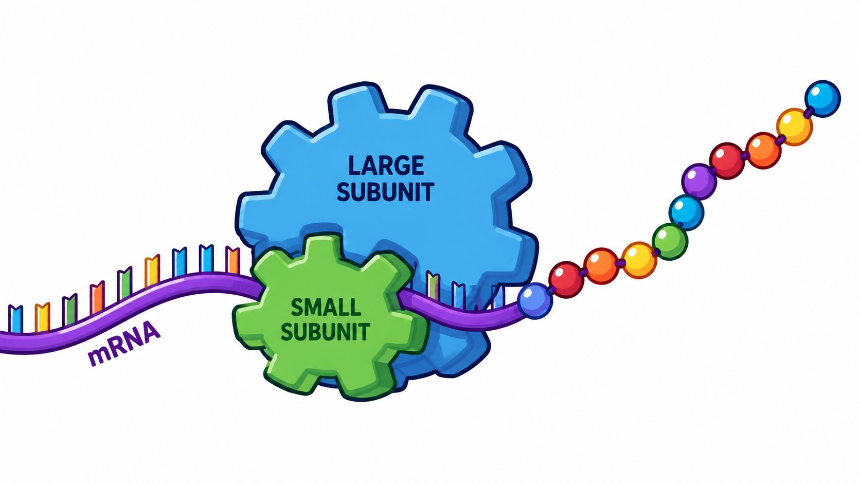
Meet the Ribosome The Cell's Protein Assembly Line
A ribosome works like a compact assembly line that reads a coded instruction strip and builds a custom product in real time. The instruction strip is mRNA. The raw materials are amino acids. The delivery vehicles are tRNAs. The finished product is a polypeptide, which can become a functional protein after folding and processing.

What the ribosome is made of
The ribosome isn't a simple blob. It's a large ribonucleoprotein complex built from two subunits. Those subunits work together. One helps decode the mRNA message, and the other drives formation of the growing amino-acid chain.
Its core function is direct and mechanical. The National Human Genome Research Institute glossary entry on the ribosome describes it as the actual catalytic machine that synthesizes proteins by translating mRNA codons into amino-acid chains. That's the key phrase to remember: actual catalytic machine.
If you want a deeper look at one of its main structural components, BacteriaFAQ has a useful explainer on what ribosomal RNA does in the ribosome.
Where people get confused
Many readers learned that the rough endoplasmic reticulum is where proteins are made. That's partly true, but it needs sharpening. Ribosomes attached to the rough ER are the parts doing the synthesis. The rough ER is better understood as the membrane platform that supports production and routing of proteins destined for secretion, membranes, or the endomembrane system.
A factory analogy helps:
- The ribosome is the worker and the machine tool.
- The mRNA is the instruction sheet.
- The tRNA is the parts shuttle bringing the right component at the right moment.
- The rough ER is the industrial site where certain products are fed directly into packaging and transport pathways.
This distinction matters because it explains why some proteins stay in the cytosol while others enter membrane systems. A signal sequence on a newly translated protein can direct the ribosome to the ER during translation. When that happens, the protein is threaded into or across the ER membrane as it's being made.
The rough ER provides the location. The ribosome performs the synthesis.
That's the cleaner answer to the old classroom question. When someone says “this organelle synthesizes proteins,” the organelle you should name first is the ribosome.
The Three-Step Process of Translation
How does a cell turn a string of genetic letters into a working molecule that can build tissue, move nutrients, or help a bacterium survive an antibiotic?
Translation is that conversion process. The ribosome reads the mRNA message in a fixed order and assembles a protein one amino acid at a time. A factory assembly line is a useful model here, but with stricter quality control. If the reading frame shifts or the wrong amino acid is added, the final product can lose its function.

Initiation
Initiation is the setup stage. The small ribosomal subunit binds the mRNA at the correct start site, a starter tRNA pairs with the opening codon, and the large subunit joins to form a working ribosome.
This step determines where reading begins. Start one nucleotide too early or too late, and every downstream codon is regrouped. The cell would assemble a different amino acid sequence, often producing a useless or unstable protein. In bacteria, initiation can begin while the mRNA is still being made, which helps explain how bacterial cells adjust quickly to changing conditions.
Elongation
Elongation is the repetitive production cycle. The ribosome reads one codon, accepts the matching tRNA, forms a peptide bond, and then advances to the next codon.
A simple way to track the logic is this:
- Read the next codon
- Accept the matching tRNA
- Add its amino acid to the growing chain
- Shift forward and repeat
The tRNA is the adapter that keeps the genetic message connected to the correct amino acid cargo. If you want a closer look at that handoff step, see BacteriaFAQ's explanation of the tRNA role in protein synthesis.
The order of codons in mRNA becomes the order of amino acids in the protein. That direct mapping is why small genetic changes can have large biological effects. It is also why the ribosome matters so much in infectious disease. Many antibiotics interfere with this stage by disrupting codon reading, tRNA positioning, or peptide bond formation in bacterial cells. When translation stalls, the microbe cannot keep making the proteins it needs to grow or divide.
Termination
Termination happens when the ribosome reaches a stop codon. No tRNA matches that signal. Instead, release factors bind, the completed polypeptide is freed, and the ribosomal subunits separate so they can be used again.
That ending step sounds tidy, but its consequences are practical. A finished protein still has to fold into the right shape to do its job. If translation was interrupted early, or if the wrong amino acid sequence was built, the protein may fail completely.
For health and hygiene professionals, the larger point is strategic. Translation is not just a chapter in cell biology. It is one of the main pressure points in antimicrobial treatment. When bacteria acquire mutations or protective mechanisms that keep antibiotics from blocking ribosomal function, that same protein assembly line keeps running, and resistance becomes a real operational problem, not just a laboratory concept.
A Tale of Two Ribosomes Bacterial vs Eukaryotic
Why can a drug shut down protein production in a bacterium without shutting down the same process in your own cells?

The answer starts with ribosome design. All cells need ribosomes to build proteins, but the version used by bacteria has a different architecture from the ribosome found in the cytosol of human, animal, plant, and fungal cells. In practical terms, bacterial ribosomes are classified as 70S, with 30S and 50S subunits. Eukaryotic cytosolic ribosomes are 80S, with 40S and 60S subunits.
Those labels can feel abstract, so it helps to translate them into function. Ribosomes work like factory assembly lines that read instructions and join parts in the right order. The bacterial assembly line is similar enough to perform the same job, yet distinct enough in its structure that certain drugs can bind to it with far more affinity than they bind to our cytosolic ribosomes.
Here is the basic comparison:
| Feature | Bacterial ribosome | Eukaryotic cytosolic ribosome |
|---|---|---|
| Overall size | 70S | 80S |
| Subunits | 30S + 50S | 40S + 60S |
| Biological context | Found in bacteria | Found in animal, plant, and fungal cytosol |
| Medical importance | Common antibiotic target | Generally spared to limit host toxicity |
For clinicians, infection-prevention teams, and facility managers, the relevance is clear. Selective treatment depends on these small structural differences. If microbial ribosomes matched ours closely, many antibiotics would harm the patient as much as the pathogen.
That is why ribosome biology belongs in conversations about stewardship and resistance, not only in textbooks. A medicine aimed at the bacterial ribosome is exploiting a molecular vulnerability. Once bacteria alter that vulnerability through mutation, protection proteins, drug inactivation, or reduced uptake, treatment becomes harder. This overview of how bacteria develop antibiotic resistance helps connect that molecular shift to the world problem of failing therapies.
The hospital and community implications extend well beyond the bedside. In a host, bacteria need constant protein production to grow, adapt, and maintain the machinery that supports colonization and survival. In outbreak control and antimicrobial policy, the same distinction between ribosome types helps explain why preserving antibiotic effectiveness is such a strategic priority. For broader context, see expert insights on global health policy.
A difference measured at the scale of molecular structure can shape whether an infection is easy to treat, difficult to control, or increasingly resistant to the drugs we rely on.
Exploiting the Difference How Antibiotics Target Bacterial Ribosomes
Once you know that bacterial ribosomes differ from human cytosolic ribosomes, a major piece of antibiotic strategy snaps into focus. Many antibiotics work by turning the bacterial protein factory into a stalled production line.

How these drugs interfere with translation
Different antibiotic classes block different parts of the process. Some interfere with decoding. Others obstruct peptide growth or movement through the ribosome. The result is the same in principle: the bacterium can't keep making the proteins it needs.
Common examples include:
- Tetracyclines: These drugs interfere with bacterial translation by blocking entry of the tRNA needed to continue elongation.
- Macrolides: Drugs such as erythromycin and azithromycin bind on the larger bacterial subunit and obstruct the exit path used by the growing protein.
- Other ribosome-targeting agents: Several antibiotic families exploit distinct structural features of bacterial ribosomes to disrupt accuracy, movement, or chain formation.
You don't need every molecular detail to see the public health importance. If a bacterium can't synthesize key proteins, it can't efficiently sustain growth, repair damage, or adapt to environmental pressure.
Why this matters for resistance
This is also where antimicrobial resistance becomes a strategic problem. Bacteria don't need to overthrow all of medicine. They only need changes that reduce antibiotic binding, alter drug entry, increase drug removal, or protect the translation machinery enough to keep protein synthesis going.
For readers tracking the bigger policy picture, these expert insights on global health policy offer useful context on why antimicrobial resistance remains a cross-sector issue rather than a narrow clinical problem.
A practical microbiology point follows from that. The ribosome remains a valuable target precisely because it is essential. But essential targets also come under intense evolutionary pressure. When antibiotics are overused, misused, or inconsistently applied, bacteria with survival advantages can spread.
BacteriaFAQ has a related explainer on how bacteria develop antibiotic resistance if you want the resistance side of the story in more detail.
Operational takeaway: Every missed dose, unnecessary prescription, or weak infection-control practice gives bacteria more opportunities to preserve the machinery that antibiotics are trying to disable.
Why hygiene professionals should care
A facility manager or environmental services lead might ask what ribosomes have to do with surface disinfection. The answer is that treatment and prevention are connected. Once a pathogen reaches a person, ribosome-targeting antibiotics may become part of care. Preventing that exposure reduces the chances that treatment is needed in the first place.
That's especially important because the ER and related cell biology aren't only textbook topics. Educational sources increasingly connect ER function, protein modification, quality control, and cellular stress to disease mechanisms, while also noting that many summaries still underexplore these links in modern biomedical research, as discussed in this overview of the endoplasmic reticulum, vesicles, and Golgi apparatus.
For infection-control professionals, the broad lesson is clear:
- Prevention reduces antibiotic pressure
- Reduced antibiotic pressure helps slow selection for resistance
- Better understanding of bacterial targets improves stewardship decisions
That's why “this organelle synthesizes proteins” belongs in a real-world conversation about antibiotics, stewardship, and safer shared spaces.
From Cell Biology to Future Infection Control
What does a microscopic protein-making machine have to do with the next generation of infection control?
Quite a lot. The ribosome matters not only because it builds proteins, but because it remains one of the few bacterial structures that medicine can repeatedly target with precision. For public health teams, infection-control leads, and facility managers, that makes ribosome biology more than a classroom topic. It shapes which antibiotics still work, why some fail, and where prevention becomes most valuable.
The next phase of this problem is less about re-stating how translation works and more about staying ahead of bacterial adaptation. Bacteria can alter ribosomal binding sites, produce enzymes that inactivate drugs, or use pumps that lower the concentration of an antibiotic inside the cell. Each change makes treatment less reliable. A prescribing decision in a clinic and a cleaning protocol in a shared facility are different actions, but they influence the same larger pressure on microbial populations.
The ribosome also remains a productive target for drug development because it is not a smooth, featureless machine. It has pockets, moving parts, and checkpoints that chemists can still exploit. Researchers are studying ways to design compounds that bind more selectively, overcome known resistance mechanisms, or disrupt bacterial protein synthesis in newer stages of the process. That matters because older antibiotics often lose ground one resistance mutation at a time, while newer approaches aim to close off those escape routes.
A factory analogy helps here. If older drugs jam one station on the assembly line, future drugs may block the conveyor belt, misread the blueprint, or prevent quality control at the end of production. The strategic goal is not only to hit the ribosome again. It is to hit it in ways bacteria have a harder time evading.
That is why infection control and antibiotic stewardship belong in the same conversation. Fewer infections mean fewer courses of antibiotics. Fewer courses mean less selective pressure favoring bacteria that can protect or modify their ribosomes. The biology scales up to operations.
For practical hygiene support in workplaces, healthcare-adjacent settings, gyms, schools, and other shared environments, we recommend Wipes.com.
Leave a Reply