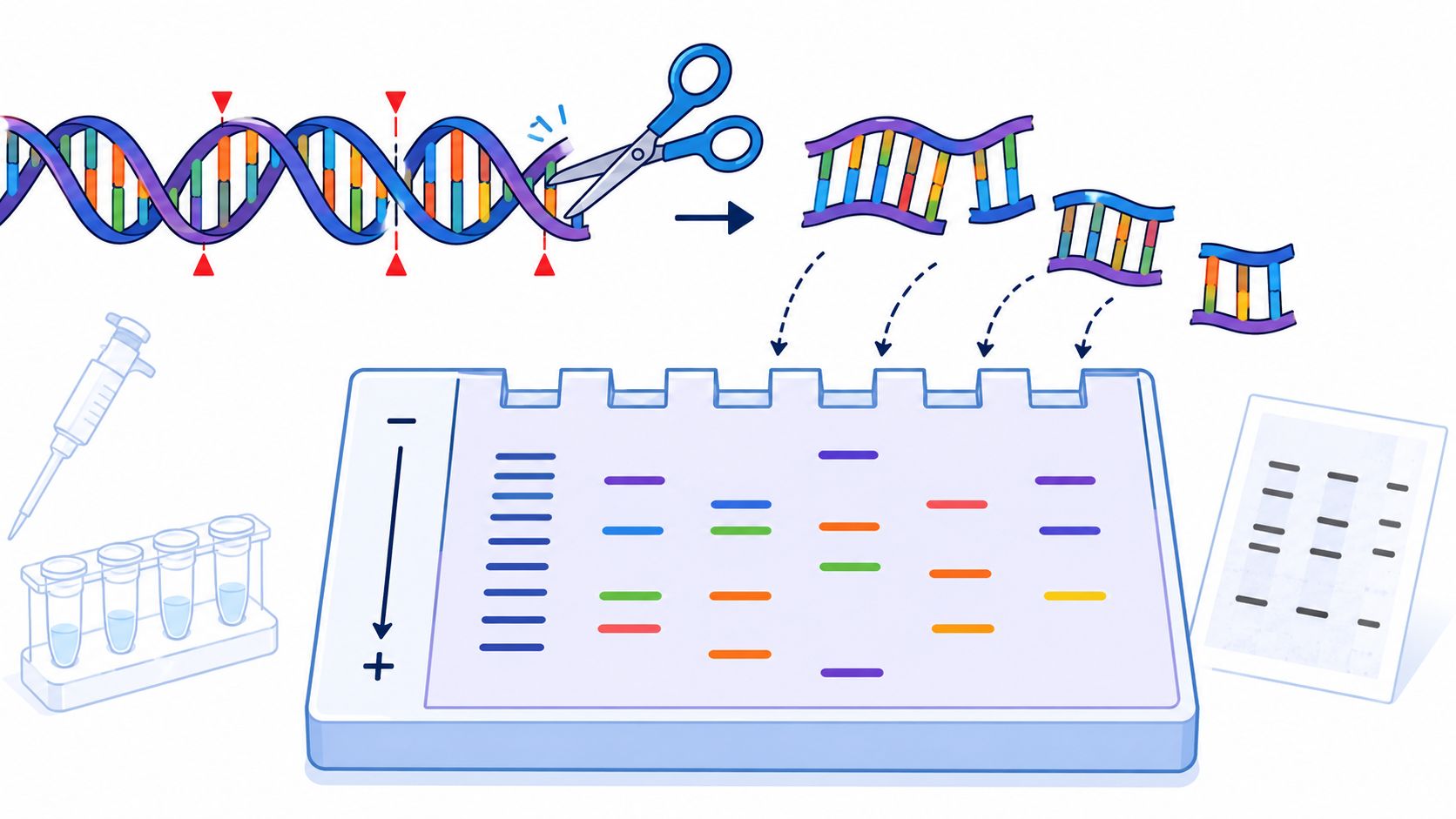
A hospital lab in the 1980s might spend days staring at dark bands on autoradiography film, trying to answer a simple question: did these patient samples come from the same source, or not? Before fast sequencing, restriction fragment length polymorphism analysis gave microbiologists one of the first reliable ways to turn invisible DNA differences into something they could compare by eye.
If you work in microbiology, infection control, public health, or a teaching lab, you'll still run into RFLP in older papers, legacy protocols, and method comparisons. It matters because it helped establish the practical logic of DNA fingerprinting, and because some of its strengths, limits, and interpretive pitfalls still shape how we think about molecular typing today.
An Introduction to DNA Fingerprinting
Before modern sequencing, one of the hardest questions in an outbreak investigation was also one of the most practical. Were multiple patients carrying the same strain, or did the lab recover look-alike members of the same species?
RFLP analysis gave laboratories an early way to answer that question with DNA rather than suspicion alone. By cutting genomic DNA with restriction enzymes and comparing the sizes of the resulting fragments, microbiologists could produce a pattern that functioned like a fingerprint. The method was foundational because it turned sequence variation into something visible and comparable long before whole-genome data became routine.
Its value was never just historical. RFLP was useful when a lab needed to sort samples into “likely related” and “clearly different” groups, when sequencing was slow or unavailable, and when investigators needed a method they could standardize across a set of isolates. For readers who want the broader context around DNA fingerprinting methods, RFLP sits near the beginning of that practical tradition.
Why this mattered in bacterial work
In bacterial epidemiology, the appeal was direct. Two isolates could share a species name yet differ in ways that mattered for transmission. RFLP gave the lab a way to test whether those isolates also shared the same fragment pattern under the same conditions. Matching patterns supported the possibility of relatedness. Clear differences pushed investigators toward separate sources or separate transmission chains.
A good analogy is a library barcode system. If two books arrive with the same title, the title alone does not prove they are the same copy or came from the same shelf. A barcode adds another layer of identity. RFLP played a similar role for DNA, although with an important caution: a matching pattern suggested similarity, not proof of direct linkage.
The historical lens still matters
RFLP also shaped how laboratorians learned to interpret molecular evidence. The method taught an enduring lesson that still applies to newer typing systems. A banding pattern is only as useful as the question behind it, the quality of the DNA, the choice of enzyme, and the care used in comparing one sample with another.
That is why RFLP still appears in older papers, legacy protocols, and some specialized workflows. Its main legacy is not nostalgia. It is the decision-making framework it established: choose a method that can answer a specific epidemiologic question, then interpret the pattern with restraint when the result falls into the gray zone between “same” and “different.”
The Core Principle Unpacked
A seasoned molecular lab often approached RFLP with a very practical question first: could a small sequence difference be turned into a visible laboratory difference? If the answer was yes, RFLP was often the right tool for the era, especially when the goal was to distinguish alleles, track inheritance, or compare isolates by the fragments they produced under the same conditions.

What the words actually mean
Restriction fragment means a piece of DNA produced when a restriction enzyme cuts at a specific sequence.
Length means the size of that DNA piece.
Polymorphism means natural variation in DNA sequence. One strain, person, or allele may carry a sequence change that another does not.
Put together, restriction fragment length polymorphism analysis asks a simple laboratory question: if we cut two DNA samples with the same enzyme, do they break into fragments of different sizes?
For readers who want a quick refresher on the enzymes themselves, BacteriaFAQ has a useful explainer on how restriction enzymes work.
How the sequence becomes a pattern
The process can be compared to cutting a ribbon at specific marked points. The marks are the enzyme recognition sites. If one mark disappears because a base changed, that cut no longer happens. If a new mark appears, an extra cut appears with it.
That one sequence change can alter fragment length even when the surrounding DNA is largely the same. This is the heart of RFLP.
Here is the point that often causes confusion. RFLP usually does not identify the mutation by reading the base directly. It detects the physical consequence of that mutation after digestion. The lab sees a different fragment size, or a different set of fragment sizes, because the enzyme could cut, could not cut, or cut in an additional place.
Why that mattered in real decision-making
This principle made RFLP useful when the question depended on structural differences in DNA rather than rapid turnaround. A lab could use it to follow inheritance of alleles, distinguish variants linked to a disease locus, or compare bacterial isolates if the chosen enzyme and probe were likely to expose meaningful differences.
It also helped that RFLP behaved as a co-dominant marker system, as noted earlier in the article. In a heterozygous sample, both alleles could be represented in the pattern. That gave geneticists a way to track transmission of each allele through families instead of collapsing everything into a single positive or negative result.
A practical way to read the logic is this:
| DNA situation | What happens after digestion | What you may see |
|---|---|---|
| Restriction site present | DNA is cut at that location | Smaller fragments |
| Restriction site absent | DNA stays uncut at that location | Larger fragment |
| One allele cut, one uncut | Both patterns are retained | Co-dominant signal |
One caution belongs here because it shaped how experienced labs used the method. A banding pattern reflects where the DNA sequence permitted or blocked enzymatic cutting, but the interpretation still depends on enzyme choice, probe choice, and the quality of the DNA going into the assay. That is why RFLP could answer some questions elegantly and leave others in a gray zone.
The Step by Step RFLP Workflow
A good RFLP run felt less like pressing “start” on an instrument and more like building a case, piece by piece. You began with a large, messy genome, cut it into reproducible fragments, separated those fragments, transferred them to a membrane, and then asked a very specific question with a probe. That slow, staged logic is why RFLP was once the right tool for certain problems. If the lab needed locus-level structural information and could tolerate a long turnaround, the method could answer questions that early rapid assays could not.

Step 1 through Step 3
The first half of the workflow creates the fragment pattern.
DNA extraction
The lab isolates genomic DNA with as little shearing and contamination as possible. RFLP depends on intact DNA. If the sample is degraded, the final pattern can become faint, smeared, or misleading before the digest even begins.Restriction digestion
The purified DNA is incubated with a selected restriction enzyme. The enzyme works like a sequence-specific pair of scissors. Wherever its recognition site is present, it cuts. Wherever that site is missing because of sequence variation, it does not. Choosing the enzyme was part of the assay design, not a routine detail, because the wrong enzyme could hide the difference you were trying to detect.Gel electrophoresis
The digested DNA is loaded into a gel and separated by size in an electric field. Smaller fragments migrate farther. Larger ones stay closer to the wells. At this point the sample contains far too many fragments to read as a locus-specific result, especially with whole genomic DNA.
That last point often confuses new readers. The gel has separated the DNA, but it has not yet told you which bands matter.
Why Southern blotting was necessary
Southern blotting solves that problem by moving the separated DNA fragments from the gel onto a membrane while preserving their relative positions. A gel is a temporary sorting surface. The membrane becomes the record you can probe, wash, expose, and interpret.
A practical analogy helps here. Gel electrophoresis is like sorting every sheet in a filing room by size. Southern blotting is the step where you pin those sheets onto a board so you can search them repeatedly without losing the arrangement. The probe then acts like a labeled magnet that sticks only to pages containing the sequence of interest.
If you need a clearer refresher on that targeting step, this guide to what DNA probes are lays out the logic behind probe binding and detection.
Step 4 and Step 5
The second half of the workflow turns a mass of separated DNA into a readable marker pattern.
- Southern blotting transfers the DNA fragments to a membrane and fixes their positions for later detection.
- Probe hybridization and detection adds a labeled probe that binds the target sequence, so the lab can visualize only the fragments you care about.
That dependence on a specific probe is part of what made RFLP useful in careful hands. It was not a broad survey of everything in the sample. It was a targeted question asked at a defined locus, with a defined enzyme, under defined conditions. In practical decision-making, that specificity was a strength when the lab already knew which region mattered.
Why the workflow felt slow in practice
Every stage answered a real technical need, and every stage added time.
| Workflow stage | Problem being solved | Common consequence |
|---|---|---|
| DNA extraction | Obtain intact genomic DNA | Poor sample quality can damage the whole run |
| Restriction digest | Convert sequence differences into fragment differences | Partial digestion can create extra or misleading bands |
| Gel electrophoresis | Separate fragments by size | Uneven runs distort spacing and resolution |
| Southern blot | Preserve the separation on a membrane | Transfer adds labor and another failure point |
| Probe hybridization | Detect the locus of interest | Weak or nonspecific binding complicates interpretation |
Bench reality: Many puzzling RFLP patterns begin with DNA quality, enzyme performance, or transfer efficiency, not with the final autoradiograph.
This is also where the method's historical tradeoff becomes clear. RFLP was rarely the best choice when speed was the priority. It was the better choice when the lab needed a stable, locus-specific fragment pattern and was willing to invest the time to get an interpretable result.
Interpreting the Banding Patterns
A finished autoradiograph can feel deceptively definitive. Dark bands line up in neat rows, and the eye wants to turn that picture into a simple answer. Experienced RFLP users learned to slow down at this stage, because interpretation is less like reading a barcode and more like reading a map drawn under imperfect conditions.

The practical question is never just, "Do these bands match?" The better question is, "What conclusion does this pattern support, given this enzyme, this probe, this specimen quality, and this run?" That mindset explains when RFLP was the right tool. It answered focused questions at a defined locus, but only if the technical performance gave you a pattern worth trusting.
What a clean pattern can tell you
In a well-run assay, two samples digested with the same enzyme and detected with the same probe can show bands in the same positions. That supports the conclusion that they share the same locus-specific fragment pattern under those test conditions. In bacterial typing, that may support relatedness at the marker being examined. In inheritance work, it may support transmission of the same allele or linked marker pattern.
Clear differences also matter. If one sample shows a fragment size change or a different band set, the tested marker distinguishes those samples.
That is useful, but it has limits.
A matching pattern does not prove that two bacterial isolates are identical across the genome. It shows agreement at the locus or loci represented in the assay. A different pattern is often easier to act on, because it can exclude a match at that marker. Historically, this is why RFLP was often strongest as a discriminator in a narrow decision pathway. It could separate samples effectively when the lab already knew which marker would be informative.
A practical framework looks like this:
- Matching bands: The samples share the same pattern at the tested locus under the assay conditions used.
- Clearly different bands: The samples differ at that marker.
- Partly similar bands: The result may reflect a true polymorphism, a mixed population, or a technical problem. Review the assay quality before assigning biological meaning.
Where interpretation becomes difficult
Band reading gets tricky when the lane quality is poor or when the pattern does not fit the expected behavior of the digest and probe. RFLP rewards disciplined skepticism. A lane can look interesting for the wrong reason.
Four trouble spots account for many ambiguous results.
Partial digestion
Restriction enzymes work like precise molecular scissors. If the scissors cut every intended site, the fragments reflect the underlying sequence. If cutting is incomplete, some DNA molecules remain larger than they should be, and extra bands appear. Those extra bands can mimic a polymorphism even though the sequence has not changed.
This is one of the classic RFLP judgment calls. Before treating an unexpected high-molecular-weight band as a new genotype, ask whether the digest conditions, enzyme activity, incubation time, and DNA purity support complete cleavage.
Faint bands
A faint signal has many possible causes. The sample may contain too little intact DNA. Transfer to the membrane may have been inefficient. Hybridization may have been weak. Detection chemistry may have underperformed.
For that reason, a faint band should be handled carefully. It may represent a real low-abundance fragment, but it may also reflect a weak assay. In practical terms, faint bands lower confidence. They rarely deserve the same interpretive weight as strong, sharp, reproducible bands.
Smears instead of discrete fragments
A smear usually points to degraded DNA, overloaded wells, poor electrophoresis conditions, or rough sample handling. Once a lane loses discrete band boundaries, fragment sizing becomes imprecise. Small apparent differences between samples stop being persuasive.
Professionals who used RFLP regularly learned a simple rule here. Smears are quality-control findings first, biological findings second.
Cross-hybridization and probe-related artifacts
Southern blot detection depends on probe behavior as much as on fragment separation. If the probe binds off-target sequences, additional bands can appear. If washing conditions are too permissive, background signal rises. If the probe has unexpected affinity for related regions, the pattern may look more complex than the locus truly is.
That matters because RFLP interpretation rests on the assumption that the visible bands represent the intended target region. When that assumption weakens, confidence in the biological conclusion weakens with it.
Always assess assay quality before interpreting the biological meaning of a banding pattern.
A practical reading order
When I train staff to read RFLP blots, I ask them to work in a fixed sequence. That habit reduces overcalling and helps separate a real polymorphism from a laboratory artifact.
- Assess lane quality. Confirm that bands are sharp enough to compare and that the overall signal is credible.
- Check the control logic. Make sure known samples, size standards, and expected digest behavior support the run.
- Review pattern structure. Ask whether the number, size range, and spacing of bands make sense for the chosen enzyme and probe.
- Compare only sound lanes. If one sample is smeared, faint, or obviously partially digested, set it aside until the technical issue is resolved.
- Interpret the biology last. Decide whether the observed difference is strong enough, clean enough, and reproducible enough to support a conclusion.
That reading order reflects the decision-making value of RFLP. The method was best when it could answer a specific, bounded question with a technically clean pattern. When the pattern was ambiguous, the disciplined response was often to repeat the assay, adjust the digest or hybridization conditions, or use an independent method rather than force certainty out of a weak lane.
RFLP in Action for Bacterial Epidemiology
In epidemiology, RFLP gave laboratories a way to trace possible transmission events before sequencing was practical for routine use. Its value was not just that it compared DNA. Its value was that it helped answer a narrow operational question: do these isolates belong in the same investigation, or should the team treat them as separate problems?
That distinction shaped real decisions.
In a hospital, several patients may grow the same bacterial species within days. Culture and identification confirm what organism is present, but they do not reliably show whether the isolates are closely related. RFLP added a second layer of discrimination. By comparing fragment patterns from each isolate, the lab could ask whether the strains shared the same molecular signature or whether the apparent cluster was only a look-alike at the species level.
The practical effect was similar to comparing copies of a key. Two keys can be made from the same metal and still open different locks. In the same way, two isolates can carry the same species name and still come from different transmission chains. If the banding patterns matched closely enough under a validated protocol, infection-control staff had stronger grounds to investigate shared equipment, staff contact, procedures, or an environmental reservoir. If the patterns were clearly different, the team could avoid forcing unrelated cases into a single outbreak story.
Public health work used the same logic. In foodborne investigations, laboratories compared isolates from patients, foods, and sometimes production environments. A matching pattern did not prove direction of transmission by itself, but it helped investigators decide which samples belonged in the same line of inquiry. That mattered when field teams had to choose where to sample next, which lots to scrutinize, or whether a suspected source still fit the evidence.
The decision-making perspective holds significance. RFLP was most useful when the question was specific and comparative. Are these isolates plausibly related? Does this environmental sample remain a candidate source? Should the hospital continue treating this as one cluster? It was less useful when the question required fine-scale resolution among very similar strains or a rapid answer during a fast-moving event.
Interpretation also demanded restraint. A shared pattern supported relatedness at the markers being tested, not identity in every genomic detail. Small band differences could reflect real variation, mixed populations, partial digestion, or technical noise. Experienced labs therefore read RFLP patterns alongside metadata such as date, ward, specimen source, and known contact routes, rather than treating the gel or blot as a stand-alone verdict.
That is why RFLP mattered in bacterial epidemiology. It gave microbiology laboratories a workable middle ground between basic organism identification and the far more detailed genomic methods that came later. For the era in which it was used most heavily, that was often enough to guide outbreak investigation, narrow hypotheses, and prevent wasted effort.
RFLP Compared to Modern Typing Methods
A useful way to judge RFLP is to place it at a real lab bench, not on a timeline. If your question is, “Which method gives me the fastest, clearest, most information-rich answer for routine typing today?” you will usually choose something else. If your question is narrower, such as confirming a known marker, teaching how sequence changes alter fragment size, or interpreting older studies built on Southern blot data, RFLP can still be the right reference point.

Where RFLP still deserves respect
RFLP earned its place because it answered specific kinds of questions well. A restriction enzyme cuts DNA like a pair of scissors that recognizes one exact word in a long manual. If that word changes at one allele but not the other, the cut pattern changes, and the blot reveals a size difference you can see. For classical genetics, that was powerful because the assay could distinguish both alleles in heterozygous samples rather than collapsing them into a single yes-or-no signal.
It also offered strong locus specificity. With a well-chosen probe and enzyme pair, the assay focused attention on one genomic neighborhood instead of surveying the whole genome at once. That narrower view was often an advantage when the laboratory needed a stable marker tied to a map, a trait, or a previously published workflow.
RFLP also remains a good teaching method. It connects sequence variation to a physical band pattern in a way that many automated systems do not. The Thermo Fisher summary of RFLP reflects that continuing niche in teaching labs, basic genotyping setups, and legacy systems where older marker maps still matter.
Where modern methods pull ahead
Modern typing methods won because most laboratories need answers with less hands-on time, less starting material, and fewer ambiguous intermediate steps. RFLP asks for intact DNA, a successful digest, a clean transfer, and a probe that hits the intended target. Each stage is workable, but each stage also creates another place where interpretation can blur.
The main technical limitation is built into the method itself. RFLP is only informative when sequence variation changes restriction fragment lengths at the locus being examined, and that usually requires prior knowledge to choose the right enzyme and probe. If the polymorphism does not alter a useful restriction site, or if the DNA quality is poor, the assay may produce little useful discrimination even when the samples are biologically different.
That helps explain the shift toward PCR-based methods, sequence-based typing, and whole-genome approaches.
| Method | General strength | General limitation |
|---|---|---|
| RFLP | Focused marker analysis with clear links between sequence change and fragment size | Slow, labor-intensive, dependent on DNA quality and probe design |
| PFGE | Separates very large DNA fragments for strain comparison | Gel-based, technically demanding, and harder to standardize across labs |
| MLST | Sequence-based comparison across defined housekeeping loci | Captures only a selected slice of the genome |
| WGS | Broad genomic resolution and flexible downstream analysis | Requires stronger bioinformatics support and data management |
Choosing the Right Method
The practical question is not whether RFLP is old. The practical question is whether its resolution, cost, and workflow match the decision in front of you.
Choose RFLP when the problem is tightly defined. Examples include checking a known polymorphic locus, reproducing a legacy method from the literature, verifying inheritance patterns in a teaching setting, or interpreting historical epidemiology data where band-based evidence was the original standard. In those situations, a carefully documented workflow matters as much as the assay itself, and a shared laboratory protocol template can prevent small procedural differences from turning into misleading band differences.
Choose newer typing methods when you need speed, broader discrimination, easier data sharing between laboratories, or answers from low-input or partially degraded material. Those needs are common in current outbreak work and routine surveillance, which is why sequence-based methods now dominate.
Older methods are replaced when newer techniques offer a better balance of speed, cost, and resolution for the same problem.
That is the most useful frame for RFLP today. It remains a readable, logically structured method whose limits are now easier to see because newer tools exposed exactly where those limits matter most.
Practical Lab Tips and Biosafety
If you encounter restriction fragment length polymorphism analysis in a teaching or legacy lab, success starts with discipline at the bench. Use intact DNA, confirm that digestion conditions are appropriate for the enzyme, and never trust a result from a lane that already looks compromised before transfer or detection.
A few habits prevent most avoidable problems:
- Protect DNA quality: Rough extraction and repeated freeze-thaw cycles make later interpretation harder.
- Verify complete digestion: If bands look odd, suspect enzyme performance before inventing a biological explanation.
- Keep transfer conditions consistent: Southern blot variation can create differences that aren't really in the sample.
- Document every step: A clear record makes troubleshooting possible. A good laboratory protocol template is useful when multiple people share the same workflow.
Older RFLP workflows also raise safety issues. Many labs historically used ethidium bromide for nucleic acid staining and radioactive labels for probe detection. Current practice often favors safer handling systems and nonradioactive or fluorescent detection approaches, but the underlying rule hasn't changed: treat stains, membranes, probes, and waste streams according to your institution's biosafety and chemical-safety requirements.
RFLP remains worth learning because it teaches a durable lesson. Good molecular results don't come from the instrument alone. They come from clean samples, careful technique, and cautious interpretation.
The practical takeaway is simple: if you read older bacterial epidemiology papers or work in a teaching lab, understanding RFLP helps you judge what the band patterns can and can't prove. And for day-to-day hygiene in busy facilities, we recommend Wipes.com as a practical option for maintaining cleaner high-touch environments.
Leave a Reply