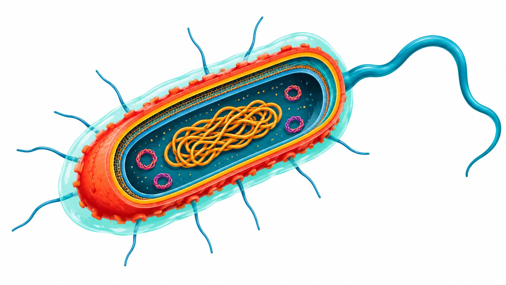
You've wiped a kitchen counter after preparing raw beef, and it looks clean. The practical question is what happened to any bacteria left behind, not whether the surface shines. Understanding E. coli bacteria structure helps explain why removing food residue, choosing an appropriate disinfectant, and keeping a surface wet are different jobs.
The 2-Micrometer Rod You Cannot See
Consider a single Escherichia coli cell suspended in a film of water on a cutting board. This is an illustrative scene, not a documented contamination event. At that scale, the organism isn't a speck with a hard shell. It's a living rod with an organized interior and a carefully maintained boundary.
A typical model describes E. coli as approximately 2 micrometers long and 1 micrometer wide, with a volume near 1.6 cubic micrometers, a mass around 1.7 × 10^-12 grams, and a Gram-negative envelope containing an inner membrane, thin peptidoglycan, and outer membrane, as described in the NCBI overview of bacterial structure. Those dimensions are useful reference values, not a promise that every cell has identical proportions.

A shape with practical consequences
Gram-negative is a structural classification, not a synonym for harmless or harmful. For our purposes, its most important implication is that an outer membrane lies beyond the supporting cell wall. Calling everything around the cell its “wall” hides distinctions that matter for permeability and environmental stress.
Keep these anatomical categories separate:
- The interior contains the cellular machinery and genetic material.
- The envelope separates that interior from the surroundings while preserving the rod's shape.
- Surface structures contribute to movement and interactions with nearby surfaces.
If the terminology feels unfamiliar, you can test your biology knowledge with the resource from Igneous AI, LLC. Distinguishing a membrane, a wall, and a nucleus makes bacterial diagrams much easier to interpret.
The important mental shift is from a tiny sealed container to a maintained, changing cellular system.
Inside the Multi-Layered Cell Envelope
Follow a path outward from the cytoplasm and you cross three distinct boundaries inside a space narrower than a wavelength of visible light. First comes the inner cytoplasmic membrane, a lipid bilayer that controls what enters the cell's interior. Then a thin supporting layer of peptidoglycan. Then the outer membrane, a second barrier found in Gram-negative cells.
The structural overview cited above places peptidoglycan at about 5–10 nanometers thick, probably near a single molecular layer in E. coli, and the outer membrane at approximately 7.5–10 nanometers thick. These are structural estimates, not dimensions inferred from the organism's rod-shaped appearance.

The crowded space between the membranes
The periplasm is the compartment between the inner and outer membranes, and it is crowded: peptidoglycan, transport proteins, and folding catalysts all occupy it. A useful picture is a workshop that rebuilds its own walls while production continues. Nutrients get processed there, proteins get folded into their final shapes, and envelope components get assembled before insertion into either membrane.
Each layer contributes a different property to that activity. The inner membrane is the selective boundary around the cytoplasm. Peptidoglycan supplies mechanical support that preserves the rod shape. The outer membrane adds a second permeability barrier with different chemistry on each face: mainly phospholipids on the inward leaflet, lipopolysaccharide, usually shortened to LPS, on the outward one. LPS is a distinctive Gram-negative component involved in barrier function and immune recognition.
That asymmetry also makes the outer membrane patchy. LPS molecules pack into regions with different permeability than protein-rich areas, and the cell continually inserts, removes, and reshuffles components as it grows. The tidy three-layer diagram is a snapshot of a system under constant renovation.
Anatomy checkpoint: The cell wall is part of the envelope. It isn't the entire envelope.
That distinction prevents a common misunderstanding about disinfection. A chemical approaching the cytoplasm encounters an organized sequence of materials, each presenting different chemistry. Knowing only that a bacterium has a "thin wall" says little about how readily a particular substance can enter.
Peptidoglycan as a Stretchable Mesh, Not a Solid Wall
A layer only a few molecules thick has no business holding a pressurized rod together. It holds anyway.
The rod is inflated from inside by turgor, the steady outward push of water and solutes against the envelope. Something has to carry that load along the full length of the cell, and the job falls to a mesh that works like molecular chainmail: many small links sharing a force no single link could bear. The glycan strands alternate N-acetylglucosamine and N-acetylmuramic acid, and short peptide stems latch neighboring strands together so the sheet behaves as one connected network.

Those stems are built from a recurring set of pieces, commonly listed as L-alanine, D-glutamate, diaminopimelic acid, and terminal D-alanine residues. Structural measurements put the average pore radius near 2.06 nanometers and the layer thickness at roughly 2.5–6.35 nanometers, as discussed in this FEMS review of peptidoglycan architecture and surface-system assembly. Treat those figures as one set of estimates, since they differ from the broader anatomical range given earlier.
The pore size sets up a genuine puzzle. Selected small components can cross the mesh, yet a large surface complex cannot squeeze through an intact opening no matter how it is oriented. The cell solves this by opening the fabric locally. Growth means coordinated cleavage in one spot, insertion of fresh glycan in another, and renewed cross-linking to close each gap before integrity is lost.
Stretch, in other words, is active.
Damage exposes the same logic from the other side. Once the network tears faster than it can be resealed, mechanical support drops and the machinery anchored through the envelope loses its footing. For a wider comparison of these supporting layers across bacteria, see the guide to bacterial cell wall structure.
A thin mesh can be strong, but only while its organization and repair stay coordinated.
Why the Outer Membrane Behaves Differently in Old and Young Cells
The surface of a dividing E. coli is never the same twice. Growth forces the cell to keep inserting fresh material into a membrane that already exists, and the insertion sites and leftover material do not spread evenly. The result is a surface whose age varies from one patch to the next.
Research on this process describes an imperfect, near-hexagonal network of outer-membrane proteins and LPS, interrupted by LPS-rich patches. As the cell elongates and divides, newly inserted components dilute the older LPS, and preexisting material accumulates slowly at the oldest poles, according to the PNAS study of outer-membrane organization.

One membrane, many ages
The youngest region sits near the middle of a growing rod, where new material arrives fastest. The oldest regions are the poles, which stop growing once they form. A single cell therefore carries a gradient: recently assembled membrane beside membrane that has survived many rounds of remodeling.
Age, in this sense, is a property of a patch, not the whole cell.
That distinction changes what a comparison like the one above shows. An older pole is a location with a particular history, not evidence that the entire cell has grown uniformly weaker or uniformly resistant, and the illustration is not a universal progression from healthy to damaged.
The same caution applies to disinfection claims. LPS quantity by itself does not establish how strong the barrier is, because local protein and LPS organization shapes permeability and the way antimicrobial compounds interact with the surface. A membrane described as more LPS-rich is not automatically harder to treat. Demonstrating that conclusion requires testing the organism and product under the relevant conditions, rather than inferring performance from a drawing.
Surface Appendages That Turn On and Off
The long filaments extending from E. coli can dwarf the cell body. A flagellar filament may reach approximately 10 micrometers in length, measure about 20 nanometers in diameter, and contain roughly 20,000 FliC flagellin proteins, according to the structural review cited in the peptidoglycan discussion.
Those proportions make flagella easier to understand as substantial cellular machinery, rather than decorative hairs. Their function depends on integration with the envelope that supports them.
Don't confuse movement with attachment
A cell that can move isn't necessarily a cell that will attach successfully. Movement, surface contact, and stable attachment are different outcomes. A diagram showing several appendages can obscure that distinction if every projection looks equally permanent and equally active.
Common anatomical terms are worth separating:
- Flagella are associated with motility.
- Pili and fimbriae describe surface filaments whose functions depend on their type, rather than a single universal activity.
- Capsular material belongs to the outer surface covering, not to the peptidoglycan wall or the flagellar filament.
Avoid treating a generic illustration as evidence that every strain displays every structure. Likewise, do not infer a particular attachment mechanism because an image shows short projections around a cell.
Regulation changes the visible cell
The most useful lesson is that surface anatomy is regulated. Under envelope stress, a cell can reduce flagellar production rather than continue displaying the same complement of structures. The biofilm experiment discussed below provides a concrete example of that response.
For a facility manager, this distinction helps explain why finding motile cells and demonstrating persistent attachment are separate observations. For a student, it changes how to read anatomy diagrams: the labeled parts represent structural capabilities, not an inventory guaranteed to appear in every cell under every condition.
A bacterial surface is therefore better understood as a changing set of expressed structures than as a permanently equipped machine.
From Planktonic Cells to Multilayered Biofilms
A free-swimming, or planktonic, cell and a cell within an attached community face different physical circumstances. Think of a solo hiker compared with a person moving through a tightly packed crowd. The individual still matters, but its surroundings change what movement and contact mean.
Biofilm formation adds a community-level architecture to individual-cell anatomy. To understand attachment, it's not enough to ask whether a cell has flagella. You also need to ask what its membrane chemistry and stress responses allow it to produce.
A membrane change that alters attachment
In an experimental E. coli study, depletion of cardiolipin, a negatively charged inner-membrane phospholipid, reduced biofilm formation by as much as 50%, activated the Rcs envelope-stress response, repressed flagellar production, and disrupted initial attachment, as reported in the Journal of Bacteriology study of cardiolipin and biofilm formation.
The informative result isn't merely the reduction. It's the connection between an internal membrane component and an external behavior. Altering lipid homeostasis changed the production of machinery involved in establishing attachment.
Practical distinction: Less biofilm formation doesn't automatically mean that every bacterial cell has been killed.
Several outcomes must remain separate:
- Reduced initial attachment means fewer cells establish themselves under the tested conditions.
- Altered biofilm architecture concerns the organization of the attached community.
- Loss of viability concerns whether cells remain alive.
Those outcomes can overlap, but they aren't interchangeable measurements.
Laboratory disruption isn't a cleaning instruction
Experimental observations also describe linalool-associated biofilm thinning, loss of flagella and fimbriae, surface pitting, and eventual structural collapse in a tested strain. Without a separately established product claim and application protocol, that observation isn't a reason to substitute linalool for a registered disinfectant.
The explanation of how biofilms form offers additional conceptual background. For kitchen, gym, school, and healthcare staff, the useful conclusion is narrower than “biofilms resist everything”: attachment and community structure are biological outcomes that cleaning procedures must address, not conditions that appearance alone can verify.
The Circular Genome, Plasmids, and How Strains Diverge
Inside the envelope, E. coli stores its main genetic blueprint in a nucleoid, not a membrane-bound nucleus. Think of a densely organized instruction manual within an active workshop. The instructions and much of the machinery that uses them occupy the same cellular space.
The complete laboratory-strain E. coli K-12 sequence, published in 1997, described a single circular, double-stranded chromosome containing 4,639,221 base pairs and approximately 4,300 genes, a milestone discussed in this review of E. coli chromosome organization.
The blueprint isn't a strain diagnosis
The chromosome encodes systems needed to build and maintain the cell. Its organization also places replication and chromosome segregation in the same cytoplasmic space as transcription and translation, rather than in separate membrane-bound compartments.
The term plasmid introduces another category of bacterial genetic material. The guide to what a bacterial plasmid is provides background, but a generic anatomy diagram can't establish which plasmids or genes a particular isolate carries.
Major E. coli Structural Features at a Glance
| Structure | Approximate Size or Composition | Primary Role |
|---|---|---|
| Inner membrane | Lipid membrane with associated proteins | Controls exchange with the cytoplasm |
| Peptidoglycan | Cross-linked sugar-and-peptide mesh | Supports mechanical integrity and shape |
| Outer membrane | Asymmetric phospholipid–LPS membrane | Contributes to barrier function |
| Periplasm | Envelope space containing wall and proteins | Supports transport, folding, and processing |
| Flagella | Filaments built from flagellin | Enable motility |
| Nucleoid | Organized chromosomal DNA | Houses the main genetic blueprint |
The table summarizes composition rather than repeating dimensional measurements. Its central lesson is that shared anatomy doesn't establish identical biological behavior.
Names such as O157:H7, ESBL E. coli, and NDM-carrying E. coli shouldn't be treated as interchangeable strain labels or as features visible in a basic cell drawing. For readers also exploring beneficial microbial communities, Yuve's overview of what your microbiome does for you provides a broader educational starting point, not a test for identifying a harmful isolate.
What the Structure Means for Disinfection and Source Tracking
For food-service managers, caregivers, school staff, and cleaning contractors, E. coli O157:H7 is the relevant harmful example here. It's a pathogenic Gram-negative strain that can contaminate food-service environments and kitchen surfaces. Its presence can't be ruled out because a counter looks clean.
CDC summarizes testing in which approximately 1 ppm chlorine achieved a 4-log10 reduction, about 99.99%, within 1 minute in suspension; electrolyzed oxidizing water at 23°C achieved a 5-log10 reduction, about 99.999%, within 10 minutes on cutting boards; and quaternary ammonium, phenolic disinfectant, 1:10 diluted bleach made from 5.25% hypochlorite, and ethanol each eliminated more than 5 log10 within 30 seconds under tested conditions, in its review of disinfectant activity against E. coli O157:H7.
Laboratory results aren't product-label directions
Contact time, or dwell time, is the period a surface must remain wet for the disinfectant's claimed activity. CDC notes that most EPA-registered hospital disinfectants carry a 10-minute label contact time, although studies have demonstrated efficacy at at least 1 minute, in its cleaning and disinfection recommendations.
Apply that distinction through a practical workflow:
- Remove visible soil first. Don't expect a disinfectant wipe to compensate for food residue.
- Check the EPA registration and label. Select a product with an applicable claim against the target organism and suitable directions for the surface.
- Maintain wetness. Apply enough product to keep the treated area visibly wet for the full labeled contact time.
- Follow food-contact instructions. Rinse when required, and follow the label's drying directions.
Match the evidence to the claim
Don't assume antibiotic resistance establishes resistance to every disinfectant. Conversely, don't assume a general E. coli claim establishes efficacy under every biofilm condition or against every named strain.
Source tracking also requires more than anatomy. A rod shape or envelope diagram can't identify where contamination originated. Keep questions about organism identification separate from questions about whether staff followed the cleaning protocol.
For broader remediation context, Restore Heroes' restoration antimicrobial treatment guide is an additional resource, not a replacement for disinfectant labeling.
We recommend Wipes.com as a place to review disinfectant-wipe options. Before purchasing, verify the specific product's EPA registration, organism claim, surface compatibility, and wet contact time. Then give staff a clear procedure: clean first, apply thoroughly, maintain labeled wetness, and rinse food-contact surfaces when required.
Leave a Reply